Table of Contents
Overview – Cerebral Blood Supply
The brain has extremely high metabolic demands, consuming 15–20% of total body energy despite comprising just 2% of body mass. This demand for oxygen and glucose is driven by neurons, which lack anaerobic capacity and rely on continuous perfusion. As a result, even short interruptions in blood flow can lead to unconsciousness or irreversible damage. Cerebral blood supply is built on redundancy, with a unique arterial anastomosis—the Circle of Willis—providing backup routes in case of vascular compromise.
Why the Brain Needs Blood
- Brain receives ~15% of cardiac output
- Neurons require constant oxygen and glucose for:
- ATP production for ion gradients
- Neurotransmitter synthesis and recycling
- No anaerobic capacity → interruption in oxygen leads to rapid loss of function
- ≈30 seconds without O₂ → unconsciousness
- Minutes without O₂ → irreversible injury
Arterial Supply – Circle of Willis
Dual-Arterial Supply System
- 2 Vertebral Arteries → unite into Basilar Artery
- 2 Internal Carotid Arteries
- These systems merge at the Circle of Willis, forming a protective arterial anastomosis
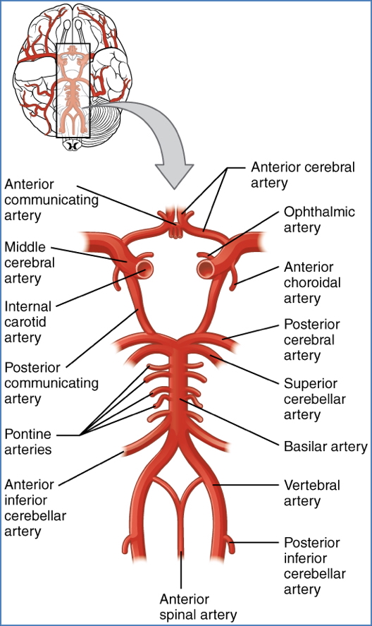
Circle of Willis – Components
- Encircles the optic chiasm, pituitary gland, and mammillary bodies
- Major branches (anterior to posterior):
- 2x Anterior Cerebral Arteries (ACA)
- 1x Anterior Communicating Artery
- 2x Internal Carotid Arteries
- 2x Middle Cerebral Arteries (MCA)
- 2x Posterior Communicating Arteries
- 2x Posterior Cerebral Arteries (PCA)
- 1x Basilar Artery
Communicating arteries are anatomically open but only functionally active when major flow is compromised (e.g. stenosis or occlusion).

Distribution of Cerebral Blood Supply
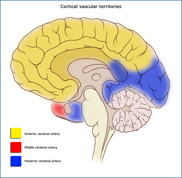
Anterior Cerebral Arteries (ACA)
- Travels along superior surface of corpus callosum
- Supplies:
- Medial frontal lobe
- Medial parietal lobe
- Corpus callosum
Middle Cerebral Arteries (MCA)
- Travels through lateral sulcus, then over lateral brain surface
- Supplies:
- Lateral frontal and parietal lobes
- Entire temporal lobe
Posterior Cerebral Arteries (PCA)
- Courses along inferior brain surface, near cerebellum
- Supplies:
- Occipital lobe (primary visual cortex)
- Posteromedial parietal lobe
- Inferior temporal lobe

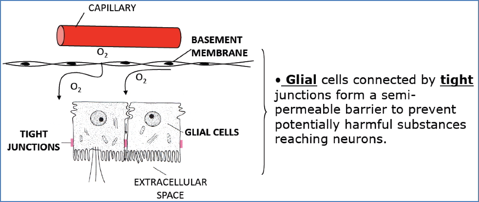
Blood–Brain Barrier (BBB)
Purpose
- Maintains a stable environment for neurons
- Limits toxin, pathogen, and ion fluctuations
Structure
- Tight Junctions between CNS capillary endothelial cells
- Blocks diffusion of most water-soluble substances
- Thick Basement Membrane
- Transport proteins for glucose, amino acids, etc.
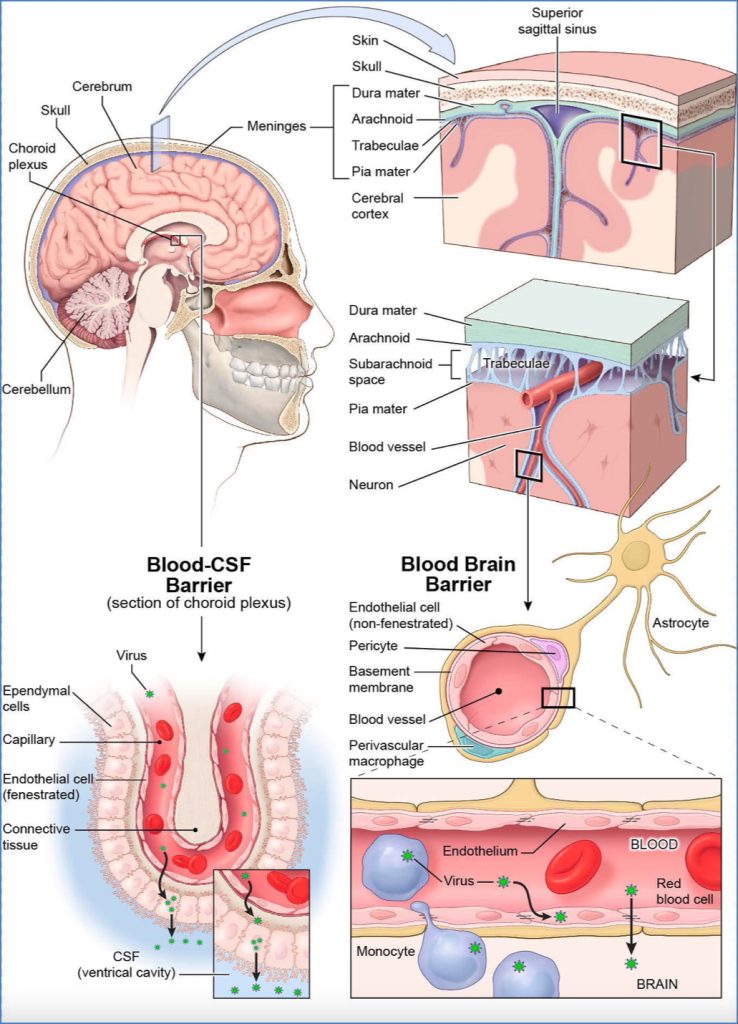
Special Considerations
- Choroid Plexuses: BBB maintained by tight junctions between ependymal cells (capillaries here are fenestrated)
- BBB is absent in:
- Hypothalamus – for hormonal sensing
- Vomiting centre (area postrema) – detects toxins in blood
Venous Drainage – Dural Sinuses
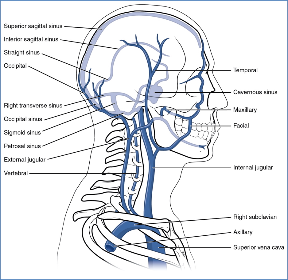
Dural Venous Sinuses
- Located within dura mater folds (not in brain tissue)
- Drain cerebral veins into internal jugular veins
Major Sinuses
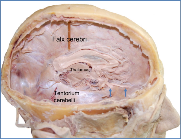
1. Falx Cerebri
- Separates cerebral hemispheres in longitudinal fissure
- Contains:
- Superior sagittal sinus
- Inferior sagittal sinus

2. Tentorium Cerebelli
- Separates cerebrum from cerebellum (transverse fissure)
- Contains:
- Left & Right transverse sinuses
Drainage Pathway
- Superior + inferior sagittal sinuses → straight sinus
- All converge into transverse sinuses → become sigmoid sinuses
- Sigmoid sinuses exit skull as internal jugular veins
Regulation of Cerebral Blood Supply
1. Autoregulation
- Maintains constant cerebral perfusion despite changes in systemic BP
- Critical in preventing stroke or ischaemia
2. Myogenic Response
- ↑Mean arterial pressure → vasoconstriction of large cerebral arteries
- ↓MAP → vasodilation to maintain perfusion
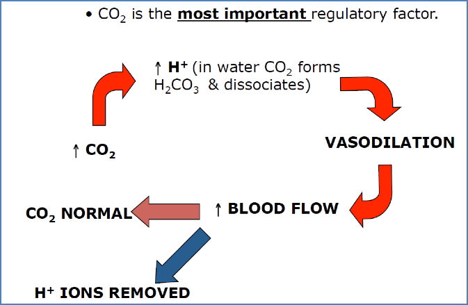
3. Metabolic Autoregulation
| Factor | Response | Effect on Flow |
| ↑CO₂ | → Vasodilation | ↑Flow |
| ↓CO₂ | → Vasoconstriction | ↓Flow |
| ↓pH (↑H⁺) | → Vasodilation | ↑Flow |
| ↑pH (↓H⁺) | → Vasoconstriction | ↓Flow |
| ↓O₂ | → Vasodilation | ↑Flow |
| ↑O₂ | → Vasoconstriction | ↓Flow |
Summary – Cerebral Blood Supply
Cerebral circulation is essential for the brain’s high metabolic demand and is protected by anatomical redundancies like the Circle of Willis. The anterior, middle, and posterior cerebral arteries provide regional cortical perfusion, while the blood–brain barrier and dural sinuses regulate and protect brain tissue. Cerebral blood flow is tightly autoregulated by myogenic and metabolic factors to prevent stroke and maintain homeostasis. For a broader context, see our Nervous System page.