Table of Contents
Overview – Skin Histology
Skin histology is essential knowledge for medical students, dermatologists, and pathologists alike. Recognising specific microscopic changes—such as acanthosis, spongiosis, or hyperkeratosis—is key to diagnosing common dermatological conditions like eczema, psoriasis, and skin cancers. This page outlines the core histopathological terms used in dermatopathology, focusing on structure–function relationships and common clinical correlations. Mastery of these concepts not only boosts exam performance but also underpins safe clinical practice.
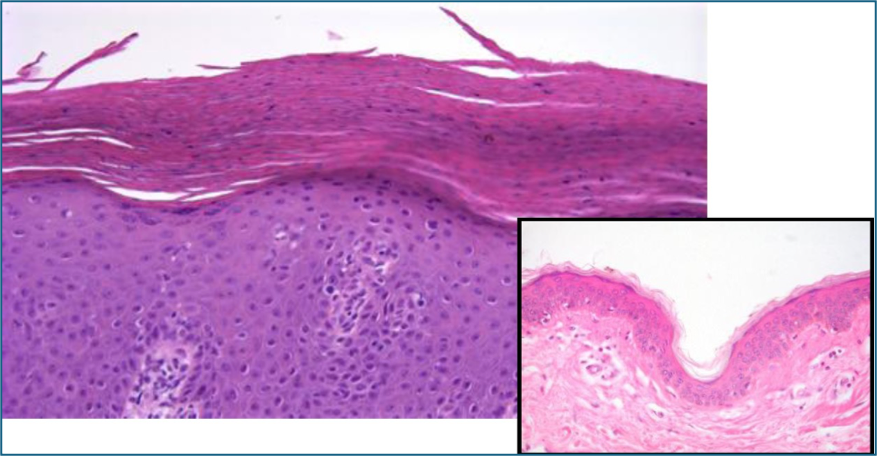
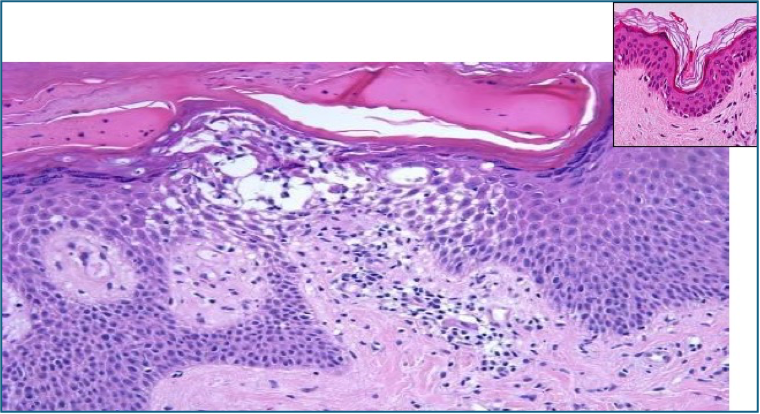
Hyperkeratosis
- = Hyperplasia of the stratum corneum (outermost skin layer)
- Often with thickening of the dermis and rete ridges
- ➝ Leads to scaling on clinical exam
- Seen in: Psoriasis, chronic eczema

Parakeratosis
- = Retention of nuclei in the stratum corneum
- Reflects rapid keratinocyte turnover without full maturation
- ➝ Causes scaling, similar to hyperkeratosis
- Note: Normal on mucosal surfaces
- Seen in: Psoriasis, actinic keratosis
Papillomatosis
- = Hyperplasia of the papillary dermis
- Creates surface projections or folds in the epidermis
- ➝ Associated with verrucous/warty growths
- Seen in: Warts, venous stasis changes, papillomas
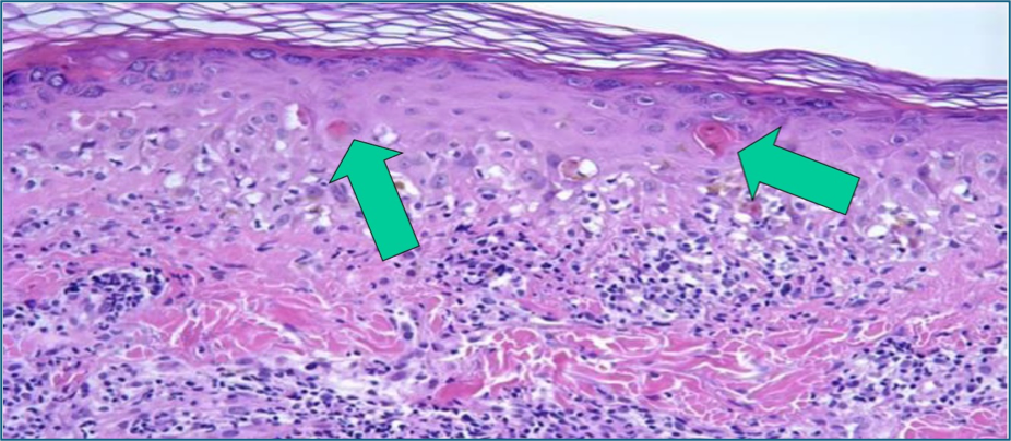
Dyskeratosis
- = Premature keratinisation of individual keratinocytes below the stratum corneum
- Often indicates malignancy or dysplasia
- May form keratin pearls or cysts
- Associated with inflammatory infiltrates
- Seen in: Squamous cell carcinoma
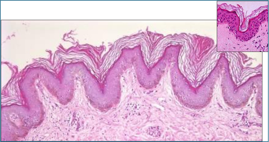
Acanthosis
- = Thickening of the epidermis due to hyperplasia of the stratum spinosum
- ➝ Causes visible thickening of skin on inspection

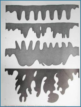
Types of Acanthosis:
- Psoriasiform (regular) – uniform elongation of rete ridges
- Irregular – variable rete ridge thickening
- Papillated – epidermis projects above skin surface
- Pseudoepitheliomatous – mimics carcinoma, invades into dermis
- Seen in: Psoriasis, chronic dermatitis
Acantholysis
- = Loss of intercellular desmosomal attachments between keratinocytes
- ➝ Leads to intraepidermal blistering
- Mechanism: Autoimmune attack (commonly against desmoglein)
- Seen in: Pemphigus vulgaris

Spongiosis
- = Interstitial oedema within the epidermis
- Results in separation of keratinocytes, but cell junctions remain intact
- Represents early inflammation or irritation
- ➝ Causes vesiculation and weeping
- Seen in: Eczema, seborrhoeic dermatitis
Summary – Skin Histology
Skin histology provides crucial insight into the pathophysiology of both inflammatory and neoplastic skin disorders. Terms like hyperkeratosis, acanthosis, and spongiosis help pathologists and clinicians correlate clinical presentations with microscopic findings. These histopathological changes underpin diagnoses ranging from eczema to malignancy. For a broader context, see our Skin & Dermatology Overview page.