Table of Contents
Overview – Cranial Nerves
Cranial nerves (CNs) are paired peripheral nerves that emerge directly from the brain and brainstem, supplying motor, sensory, and autonomic innervation to the head, neck, and beyond. While functionally similar to spinal nerves, cranial nerves are highly specialised, reflecting embryological origins such as pharyngeal arches and somite-derived musculature. This page is an overview of their functional classification, developmental context, associated ganglia, and brainstem organisation — forming the core framework for understanding the individual cranial nerves I–XII.
Structural Similarities to Spinal Nerves
- Cranial nerves resemble spinal nerves in:
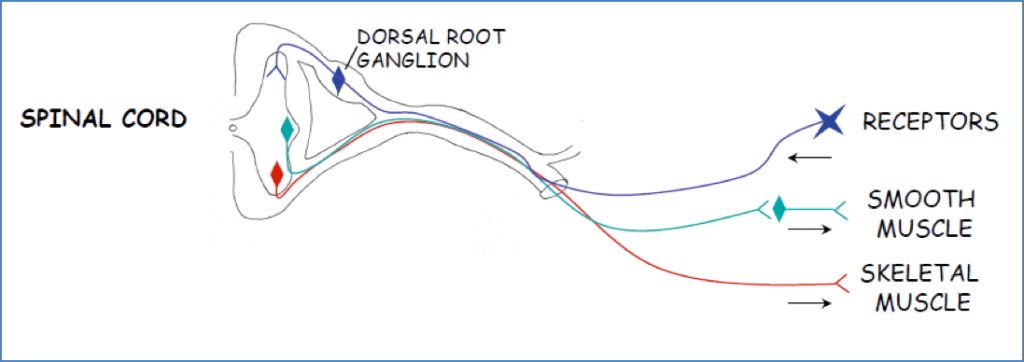
- Sensory organisation: Dendrites receive input → cell bodies in sensory ganglia → axons terminate in sensory nuclei of the brainstem
- Touch → Posterior columns
- Pain → Spinothalamic
- Proprioception → Spinocerebellar
- Somatic motor organisation: Cell bodies in motor nuclei of brainstem → axons innervate skeletal muscle
- Visceral motor organisation: Preganglionic neurons from visceral nuclei synapse in autonomic ganglia, then project to smooth muscle/glands
- Sensory organisation: Dendrites receive input → cell bodies in sensory ganglia → axons terminate in sensory nuclei of the brainstem
Functional Components of Cranial Nerves
Cranial nerves may carry one or more of five functional fibre types:
1. Voluntary (Somatic) Motor
- GSE (General Somatic Efferent): Skeletal muscle from somites (e.g. eye and tongue muscles)
- SVE (Special Visceral Efferent): Skeletal muscle from pharyngeal arches (e.g. muscles of mastication, facial expression, swallowing)
2. Involuntary (Visceral) Motor
- GVE (General Visceral Efferent): Parasympathetic supply to glands and smooth muscle (2-neuron system via ganglia)
3. Somatic Sensation
- GSA (General Somatic Afferent): Touch, pain, temperature from skin, mucosa, and other somatic structures
4. Visceral Sensation
- GVA (General Visceral Afferent): Internal organ sensory input (e.g. BP, chemoreception, GI sensation)
5. Special Sensation
- SSA/SVA (Special Somatic/Visceral Afferent): Vision, hearing, balance, taste, smell
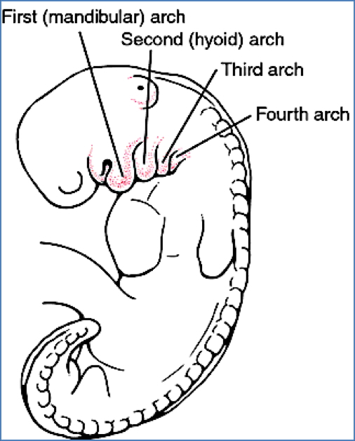
Pharyngeal Arch Associations
| Arch | Associated CN | Muscles Derived |
|---|---|---|
| 1st (Mandibular) | CN V (Trigeminal) | Muscles of mastication, anterior digastric, tensor tympani |
| 2nd (Hyoid) | CN VII (Facial) | Muscles of facial expression, posterior digastric, stylohyoid |
| 3rd | CN IX (Glossopharyngeal) | Stylopharyngeus |
| 4th | CN X (Vagus) | Cricothyroid, soft palate muscles |
| 6th | CN X (Vagus) | Intrinsic laryngeal muscles |
Note: 5th arch is transient and has no adult derivative.
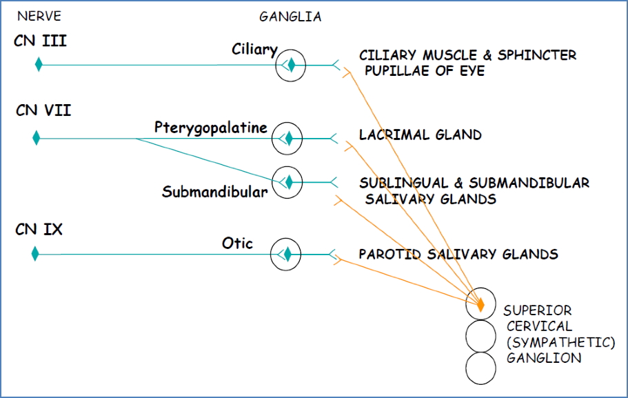
Parasympathetic Ganglia of Cranial Nerves
| CN | Ganglion | Target |
|---|---|---|
| CN III (Oculomotor) | Ciliary | Eye – pupil constriction, lens accommodation |
| CN VII (Facial) | Pterygopalatine | Lacrimal gland |
| CN VII (Facial) | Submandibular | Submandibular & sublingual salivary glands |
| CN IX (Glossopharyngeal) | Otic | Parotid salivary gland |
Sensory Ganglia of Cranial Nerves
| CN | Ganglion | Sensory Type |
|---|---|---|
| I (Olfactory) | Olfactory epithelium | Smell |
| II (Optic) | Retina | Vision |
| V (Trigeminal) | Trigeminal ganglion | Touch, pain, temperature |
| VII (Facial) | Geniculate | Taste (ant. 2/3 tongue), external ear |
| VIII (Vestibulocochlear) | Vestibular + Spiral | Balance + hearing |
| IX (Glossopharyngeal) | Inferior ganglion | Taste, BP, chemoreception |
| X (Vagus) | Superior & Inferior ganglia | Visceral sensation, taste, external ear |
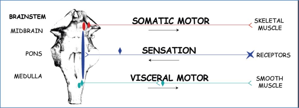
Cranial Nerve Nuclei
- CN I & II → Forebrain (not true brainstem CNs)
- CN III–XII → Brainstem
Brainstem Column Organisation (Generalised)
- Medial (Motor):
- GSE (e.g. CN III, IV, VI, XII)
- SVE (e.g. CN V, VII, IX, X)
- GVE (parasympathetic)
- Lateral (Sensory):
- SSA/SVA (e.g. CN VIII, I, II)
- GSA/GVA (e.g. CN V, IX, X)
Cranial Foramina Summary
| Foramen | Cranial Nerves |
|---|---|
| Cribriform plate | CN I |
| Optic canal | CN II |
| Superior orbital fissure | CN III, IV, V₁, VI |
| Foramen rotundum | CN V₂ |
| Foramen ovale | CN V₃ |
| Internal acoustic meatus | CN VII, VIII |
| Jugular foramen | CN IX, X, XI |
| Hypoglossal canal | CN XII |
| Foramen magnum | CN XI (spinal root enters skull) |

Summary – Cranial Nerves Overview
Cranial nerves follow the basic organisation of spinal nerves but are highly specialised for motor, sensory, and autonomic functions within the head and neck. They develop in association with somites and pharyngeal arches, carry distinct functional fibre types, and emerge through specific cranial foramina. Understanding this structural and developmental context is essential before learning each individual nerve. For individual nerve anatomy and function, see our Nervous System Overview.