Table of Contents
Overview – GI Neurovascular Supply
The gastrointestinal (GI) tract relies on a highly coordinated network of arteries, veins, and nerves to function effectively. The arterial supply corresponds closely with embryological origins (foregut, midgut, hindgut), while venous return channels nutrients to the liver via the portal system. Neural regulation involves both intrinsic plexuses and extrinsic autonomic inputs. Understanding the GI neurovascular supply is essential for interpreting abdominal pain, planning surgical procedures, and managing vascular or neuropathic GI pathologies.
Arterial Supply of the GI Tract
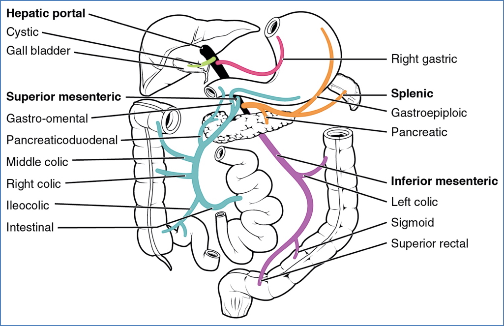
Foregut – Supplied by the Celiac Trunk
- Pharynx
- Oesophagus
- Stomach
- Proximal duodenum (upper portion)
- Liver
- Gallbladder
- Spleen
- Upper ½ of pancreas
- Respiratory tract (including lungs)
Midgut – Supplied by the Superior Mesenteric Artery (SMA)
- Lower duodenum
- Jejunum
- Ileum
- Cecum
- Appendix
- Ascending colon
- Proximal 2/3 of transverse colon
- Lower ½ of pancreas
Hindgut – Supplied by the Inferior Mesenteric Artery (IMA)
- Distal 1/3 of transverse colon
- Descending colon
- Sigmoid colon
- Rectum
- Upper anal canal
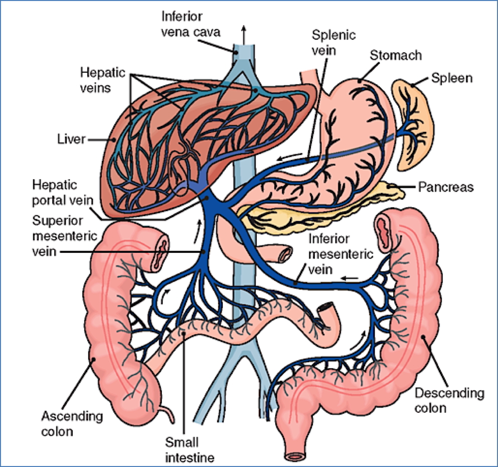
Venous Drainage of the GI Tract
Foregut
- Portal vein (main route to liver)
- Left and right gastric veins → Portal vein
- Splenic vein → Portal vein
Midgut
- Superior mesenteric vein → Portal vein
Hindgut
- Inferior mesenteric vein → Splenic vein → Portal vein
All nutrient-rich blood from the GI tract drains to the liver via the hepatic portal system for metabolic processing and detoxification.
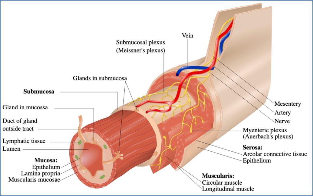
Nervous Supply of the GI Tract
Intrinsic Innervation
- Derived from two major plexuses:
- Myenteric (Auerbach’s) plexus → Between circular and longitudinal layers of muscularis externa
- Submucosal (Meissner’s) plexus → In the submucosa
- Function:
- Coordinates local motility, secretion, and blood flow
- Operates largely independently (enteric nervous system)
- Extends from oesophagus to rectum
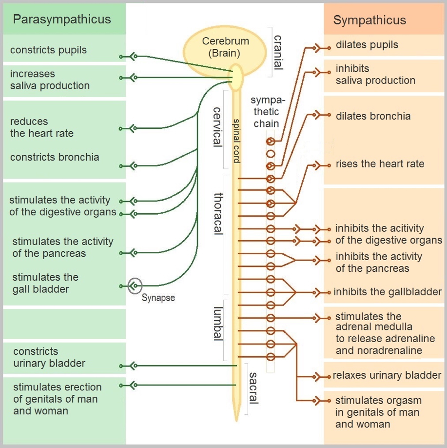
Extrinsic Innervation
Parasympathetic (rest and digest)
- Vagus nerve: Foregut and midgut
- Pelvic splanchnic nerves (S2–S4): Hindgut
Sympathetic (fight or flight)
- Thoracic splanchnic nerves (greater, lesser, least)
- Inhibit peristalsis and secretion; contract sphincters
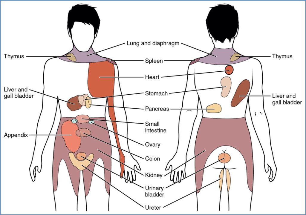
Referred Pain in GI Disorders
- GI organs often lack distinct somatic sensory innervation
- Pain from visceral structures is interpreted as arising from skin or muscle overlying the corresponding spinal segment
- This phenomenon is known as viscero-somatic convergence
- Example:
- Diaphragmatic irritation → Shoulder tip pain (C3–C5)
- Appendix inflammation → Periumbilical pain (T10)
Summary – GI Neurovascular Supply
The gastrointestinal neurovascular supply integrates embryological segmentation with vascular and neural anatomy. Arterial blood supply is divided into foregut (celiac trunk), midgut (SMA), and hindgut (IMA), while venous return enters the hepatic portal system for liver processing. Neural regulation is both intrinsic (enteric plexuses) and extrinsic (autonomic nervous system), with referred pain pathways frequently seen in clinical practice. For a broader context, see our Gastrointestinal Overview page.