Table of Contents
Overview – Liver Anatomy
The liver is the largest internal organ and a vital multifunctional gland that plays central roles in metabolism, detoxification, immunity, and bile production. Weighing around 1.5 kg, it receives dual blood supply and interfaces directly with the gastrointestinal tract via the portal venous system and biliary tree. A deep understanding of liver anatomy, structure, and function is essential for interpreting liver pathology, surgical approaches, and systemic metabolic disturbances.
Functions of the Liver
- Bile synthesis – essential for fat digestion
- Protein metabolism – synthesis, storage, degradation (including transamination)
- Carbohydrate metabolism:
- Glycogenesis: Glucose → Glycogen
- Glycogenolysis: Glycogen → Glucose
- Lipid metabolism – VLDL/HDL synthesis, cholesterol handling
- Vitamin A production & storage
- Anticoagulant synthesis – Heparin production
- Drug detoxification & activation
- Immunological function – Kupffer cells (liver macrophages)
Gross Structure of the Liver
- Size & Shape:
- ~1.5 kg, wedge-shaped
- Two main surfaces:
- Diaphragmatic surface
- Visceral surface
- Lobes:
- Right
- Left
- Quadrate
- Caudate
- Peritoneal Relationships:
- Covered by peritoneum except at the bare area
- Falciform ligament divides right and left lobes
- Ligamentum teres – remnant of fetal umbilical vein
- Lesser omentum – attaches to stomach’s lesser curvature

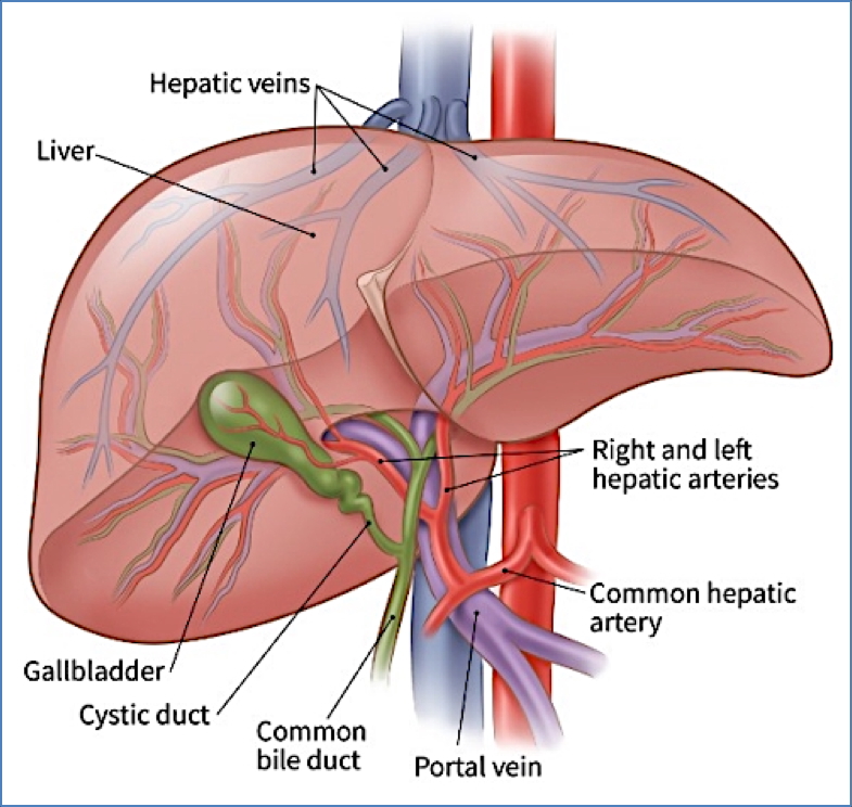
Blood Supply
- Dual Blood Supply:
- Hepatic artery (~20%) – oxygenated blood
- Portal vein (~80%) – nutrient-rich blood from GIT
- Venous Drainage:
- All liver blood drains into the inferior vena cava
The Portal Triad
- Hepatic artery – from celiac trunk
- Portal vein – drains blood from GI tract, spleen, pancreas
- Common hepatic duct – drains bile from liver
- The portal triad runs through the lesser omentum
Liver Innervation & Lymphatics
- Sympathetic: From L/R sympathetic trunks
- Parasympathetic: Via vagus nerve (Cranial Nerve X)
- Lymph Drainage:
- Celiac, hepatic, cystic, gastric, pyloric, and superior mesenteric nodes
Microscopic Structure – Liver Lobules
- Hexagonal lobules centred around a central vein
- At each corner (portal triad):
- Branch of hepatic artery
- Branch of portal vein
- Bile duct
- Zones of perfusion:
- Zone 1: Closest to portal triad – receives highest oxygen
- Zone 2: Intermediate
- Zone 3: Centrilobular zone – most vulnerable to ischaemia/toxins

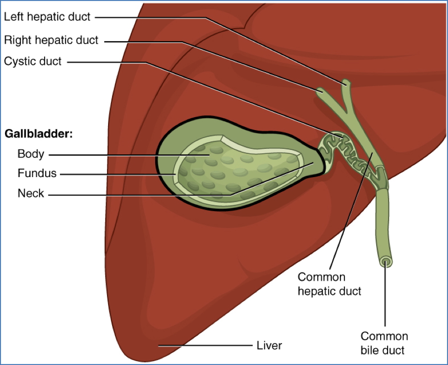
The Biliary Tree
Intrahepatic Bile Flow
- Hepatocytes → bile canaliculi → small ducts → merge into right and left hepatic ducts
Extrahepatic Pathways
- Right + Left hepatic ducts → Common hepatic duct
- Common hepatic duct branches to:
- Cystic duct → gallbladder
- Common bile duct → joins with pancreatic duct → forms hepatopancreatic ampulla (of Vater)
- Ampulla opens into duodenum via the major duodenal papilla
- Controlled by: Sphincter of Oddi

2. https://muschealth.org/medical-services/ddc/patients/digestive-diseases/pancreas/sphincter-of-oddi-dysfunction
Summary – Liver Anatomy
The liver is a highly vascularised organ critical for metabolic, immune, and digestive functions. It receives dual blood supply, processes GI-absorbed nutrients via the portal vein, and produces bile for lipid digestion. Structurally divided into lobes and microscopic lobules, its functional units are arranged around a portal triad. For a broader context, see our Gastrointestinal Overview page.