Table of Contents
Overview
Leishmaniasis is a parasitic disease caused by Leishmania species, transmitted through the bite of infected sandflies. It manifests in three major clinical forms—visceral, cutaneous, and mucocutaneous—depending on the species and immune response. Leishmaniasis remains a major neglected tropical disease, affecting millions in endemic regions, with serious morbidity and potential mortality in its visceral form.
Definition
Leishmaniasis refers to a group of diseases caused by protozoan parasites of the genus Leishmania, transmitted to humans via the bite of infected female sandflies.
Aetiology
- Pathogen: Leishmania protozoa
- Vector: Phlebotomine sandflies (ecto-parasites)
- Geographic Distribution: Predominantly tropical, subtropical, and southern European regions
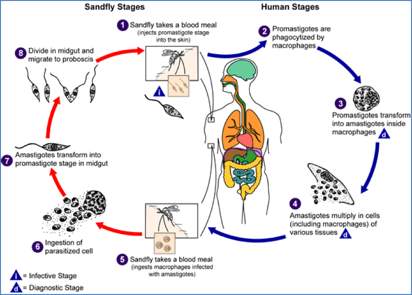
Lifecycle
- Amastigotes: Intracellular form found in humans, particularly within macrophages
- Promastigotes: Motile extracellular form found in the sandfly’s gut
- Lifecycle:
- Sandfly takes a blood meal and injects promastigotes into the human host
- Promastigotes are phagocytosed by macrophages → transform into amastigotes
- Amastigotes multiply → infect other cells
- Sandfly ingests amastigotes during next blood meal → develop into promastigotes in gut
Clinical Features
Visceral Leishmaniasis (Kala Azar)
- Systemic disease
- Classic symptoms:
- Fever
- Weight loss
- Hepatosplenomegaly (especially splenic enlargement)
- Anaemia and pancytopenia
- Hyperpigmentation (“Kala Azar” = “black fever”)
- Fatal if untreated
Cutaneous Leishmaniasis
- Most common form
- Features:
- Papules → nodular → ulcerated lesions
- Typically painless
- Can result in disfiguring scars
- Localised to skin, often self-limiting
Mucocutaneous Leishmaniasis
- Occurs when cutaneous disease spreads to nasopharyngeal mucosa
- Features:
- Destructive and disfiguring ulcers
- Involvement of nose, mouth, and throat
- Can cause facial deformities

Investigations
- Microscopy: Detection of amastigotes in tissue smears
- Culture: From aspirates or biopsies
- Serology: Useful for visceral disease
- PCR: High sensitivity for species identification
- Montenegro skin test (delayed hypersensitivity) – useful in endemic surveillance
Management
- Visceral Leishmaniasis:
- Liposomal Amphotericin B (first-line in many settings)
- Miltefosine or antimonials (in some endemic areas)
- Cutaneous/Mucocutaneous Leishmaniasis:
- May be self-resolving (mild cases)
- Local therapy: heat or cryotherapy
- Systemic therapy for severe, multiple, or mucosal involvement
- Supportive care: Manage anaemia, nutritional support, treat secondary infections
Prevention
- Avoid sandfly bites:
- Insect repellents, protective clothing
- Bed nets (especially fine mesh)
- Vector control (spraying and environmental hygiene)
- Public health education
- Surveillance and early detection in endemic zones
Summary
Leishmaniasis is a parasitic disease caused by Leishmania species and transmitted by sandflies, presenting in visceral, cutaneous, and mucocutaneous forms. Visceral leishmaniasis (Kala Azar) is potentially fatal without treatment, whereas cutaneous and mucocutaneous forms can cause significant disfigurement. Management involves antiparasitic therapy and vector control. For broader context, see our Microbiology & Public Health Overview page.