Table of Contents
Overview – Bones of the Skull
The bones of the skull form the protective casing for the brain and the structural framework of the face. They house sensory organs, provide anchor points for muscles, and contribute to both respiration and mastication. A solid understanding of skull anatomy is foundational for neuroanatomy, head trauma assessment, and surgical approaches in ENT and neurosurgery.
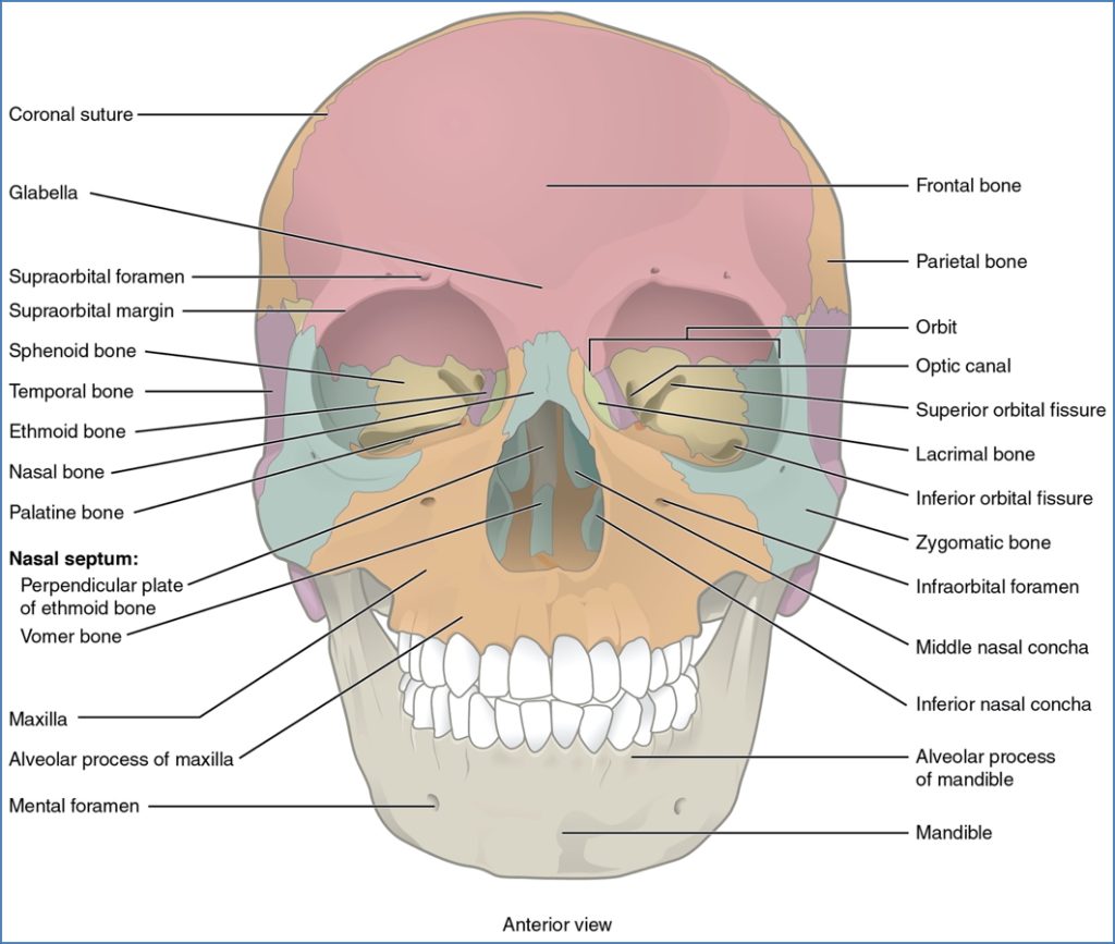
Anterior View of the Skull
The front view of the skull shows the key facial and some cranial bones.
- Frontal Bone – Forms the forehead and the superior orbital rim
- Maxilla – Upper jaw; houses upper teeth and forms part of the nasal aperture
- Mandible – Lower jaw; the only moveable skull bone
- Nasal Bones – Paired bones forming the bridge of the nose
- Zygomatic Bones – Cheekbones; form part of the lateral orbital wall
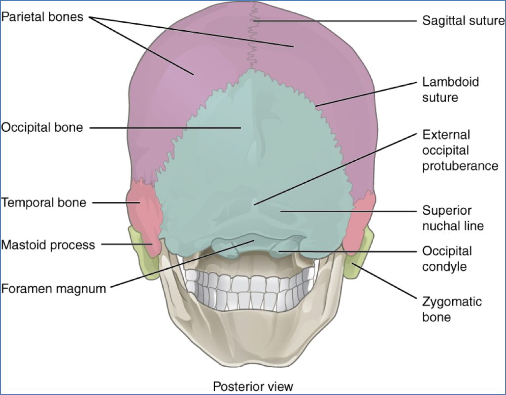
Posterior View of the Skull
Seen from behind, the skull’s contour is largely formed by the:
- Parietal Bones – Paired; form the superior and lateral cranial vault
- Occipital Bone – Forms the posterior skull and encloses the foramen magnum
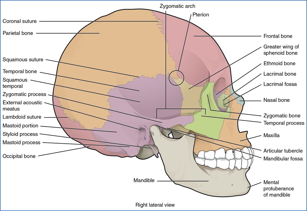
Lateral View of the Skull
The side view reveals overlapping and deeply integrated bones:
- Temporal Bone – Houses the external acoustic meatus and mastoid process
- Parietal Bone – Large, flat bone of the lateral skull
- Ethmoid Bone – Small contribution visible at the nasal region
- Frontal Bone – Extends into the superior orbital rim and forehead
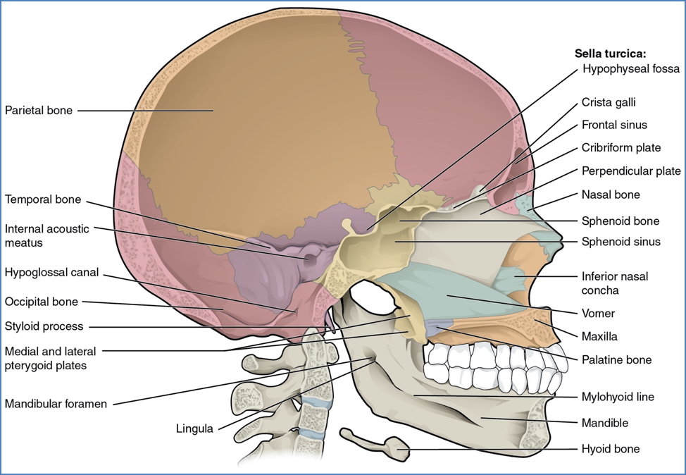
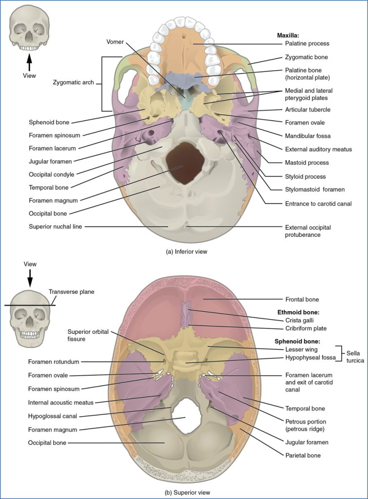
Inferior Aspect of the Skull
Seen from below, the skull base consists of bones supporting the oral and cranial cavities:
- Maxilla & Palatine Bones – Form the hard palate
- Sphenoid Bone – Butterfly-shaped; forms part of the cranial floor
- Occipital Bone – Contains the foramen magnum
- Temporal Bone – Includes the mandibular fossa for the temporomandibular joint (TMJ)
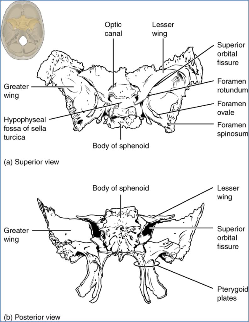
Special Focus: Sphenoid Bone
- Shape: Butterfly-shaped bone located at the base of the skull
- Articulations: Connects with nearly every other cranial bone
- Key Features:
- Sella turcica – houses the pituitary gland
- Greater and lesser wings – form parts of the cranial cavity and orbits
- Pterygoid processes – important for muscle attachment
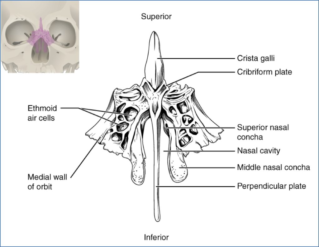
Special Focus: Ethmoid Bone
- Location: Anterior cranial base, between the orbits
- Functions: Forms part of the nasal septum, medial orbital walls, and nasal cavity roof
- Key Features:
- Cribriform plate – allows passage of olfactory nerves
- Crista galli – attachment for dura mater
- Perpendicular plate – contributes to the nasal septum
Summary – Bones of the Skull
The skull consists of multiple bones that interlock to protect the brain and support facial structure. Key bones include the frontal, parietal, temporal, occipital, maxilla, mandible, and the centrally important sphenoid and ethmoid bones. This knowledge is essential for understanding trauma, surgical planning, and neurological assessment. For a broader context, see our Musculoskeletal Overview page.