Table of Contents
Overview – Lower Limb Bones
The lower limb bones form the structural framework for weight-bearing, locomotion, and balance. Comprising the pelvic girdle, thigh, leg, and foot, they work synergistically to support body mass and enable a wide range of lower limb movements. A solid understanding of their landmarks, articulations, and muscle attachments is essential for clinical anatomy, orthopaedics, and surgical planning.
Bony Pelvis
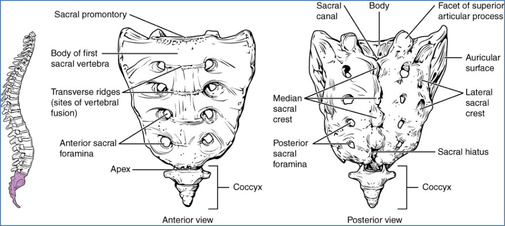
Sacrum
- Type: Irregular bone; fusion of 5 sacral vertebrae
- Landmarks:
- Transverse ridges → sites of vertebral fusion
- Anterior/Posterior sacral foramina → transmit sacral spinal rami and vessels
- Median sacral crest → fused spinous processes
- Lateral sacral crests
- Sacral canal → continuation of vertebral canal
- Sacral hiatus → inferior external opening
- Articulations:
- L5 vertebra
- Coccyx
- Hip bones (via sacroiliac joints)
- Origins/Insertions:
- Iliacus (origin)
- Gluteus maximus (origin)
- Piriformis (origin)
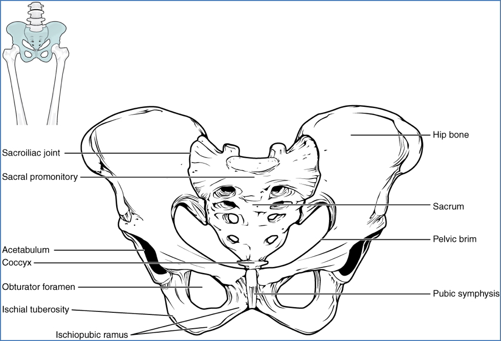
Coxal Bones (Hip Bones)
- Type: Irregular bones; fuse from ilium, ischium, and pubis in childhood
- Landmarks:
- Acetabulum → hemispherical socket for femur
- Pelvic brim
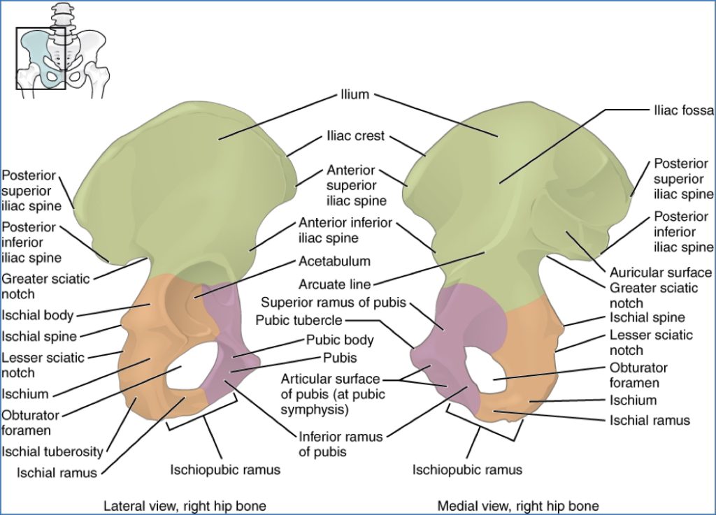
- Ilium:
- Iliac crest, ASIS, PSIS, AIIS, PIIS
- Iliac fossa
- Greater sciatic notch (passage for sciatic nerve)
- Ischium:
- Ischial tuberosity (sacrotuberous ligament attachment)
- Pubis:
- Pubic tubercle, symphysis, obturator foramen
- Subpubic angle (wider in females)
- Articulations:
- Sacrum
- Contralateral hip bone
- Femurs
Femur
- Type: Longest and strongest long bone
- Angles:
- Angle of inclination ≈ 125°
- Anteversion ≈ 10°
- Neck prone to fracture due to poor trabecular support
- Landmarks:
- Head, neck, fovea capitis
- Greater and lesser trochanters
- Intertrochanteric line (anterior) and crest (posterior)
- Gluteal tuberosity, linea aspera
- Medial/lateral epicondyles and condyles
- Intercondylar fossa
- Adductor tubercle
- Patellar surface
- Articulations:
- Acetabulum
- Tibia
- Patella
- Origins/Insertions:
- Multiple: including iliacus, gluteals, adductors, gastrocnemius, plantaris, popliteus, etc.

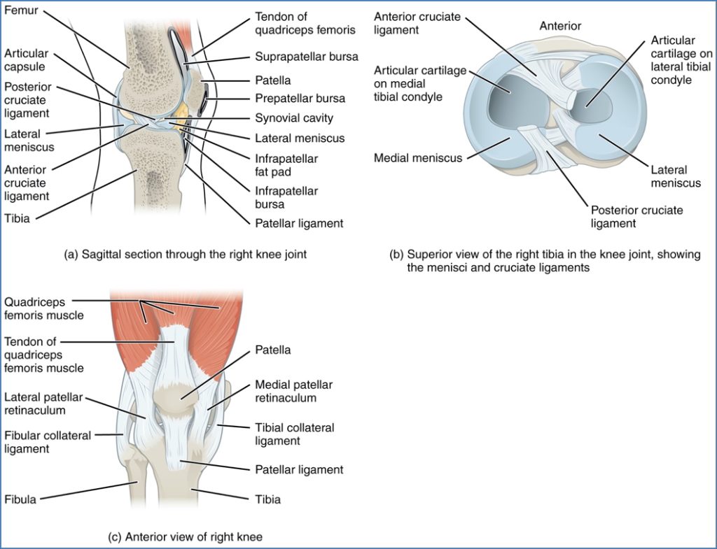
Patella
- Type: Triangular sesamoid bone
- Features:
- Enclosed in quadriceps tendon
- Improves leverage of knee extension
- Protects anterior knee
- Landmarks:
- Apex
- Medial and lateral facets
- Articulations:
- Femoral condyles (flexed knee)
- Patellar surface of femur (extended knee)
- Origins/Insertions:
- Insertion point for quadriceps via quadriceps tendon
- Patellar ligament connects to tibial tuberosity
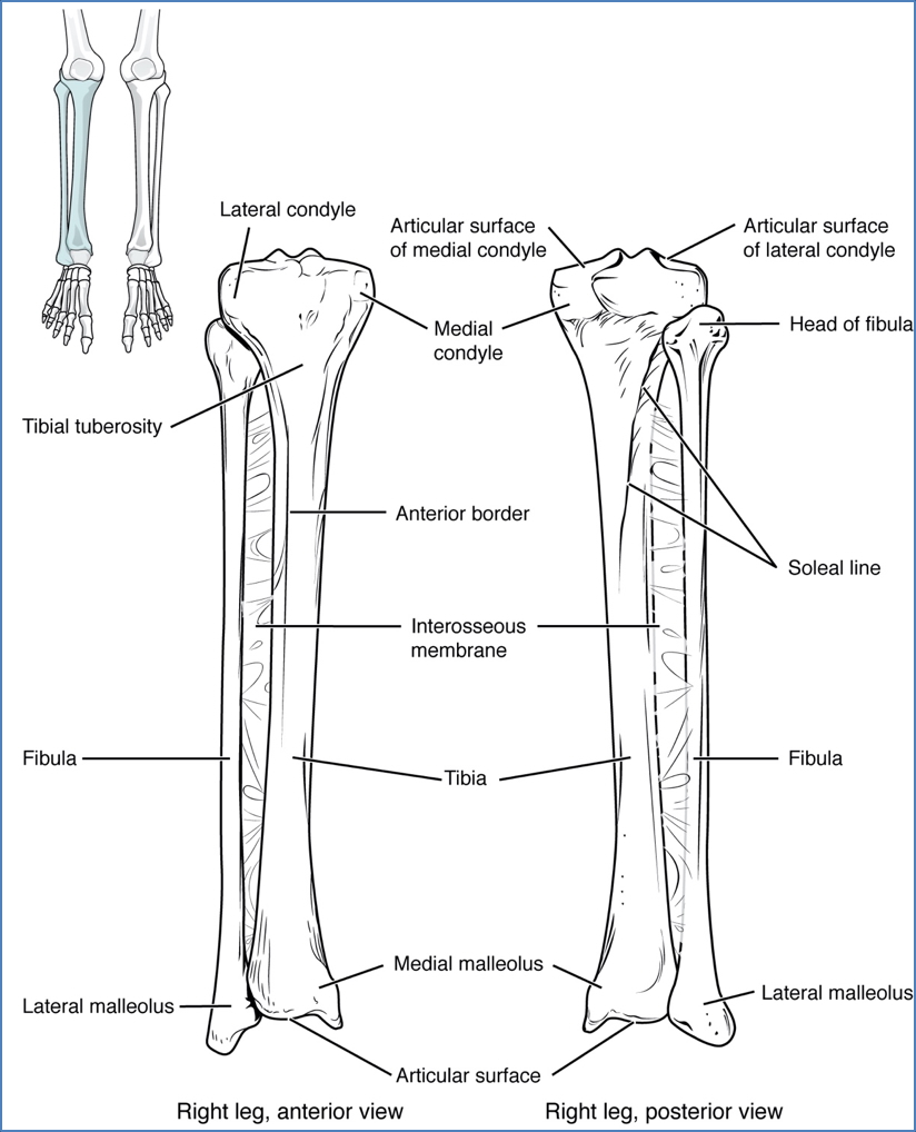
Tibia
- Type: Long bone; primary weight-bearing bone of the leg
- Landmarks:
- Medial/lateral condyles
- Intercondylar eminence & tubercles
- Tibial tuberosity
- Soleal line
- Anterior/interosseous borders
- Medial malleolus
- Fibular facet (proximal)
- Fibular notch (distal)
- Articulations:
- Femoral condyles
- Talus (ankle joint)
- Fibula (proximal & distal)
- Origins/Insertions:
- Includes: quadriceps femoris, sartorius, semitendinosus, tibialis anterior/posterior, soleus, FDL, EDL
Fibula
- Type: Slender long bone
- Features:
- Does not bear weight
- Important for muscle attachment
- Fixed – unlike radius/ulna (no supination/pronation)
- Connected to tibia via interosseous membrane
- Landmarks:
- Head, neck
- Shaft (anterior/interosseous/posterior borders)
- Lateral malleolus
- Articulations:
- Tibia (both ends)
- Talus
- Origins/Insertions:
- Includes: biceps femoris, fibularis group, tibialis posterior, flexor/extensor hallucis/digitorum longus
Bones of the Foot
- Tarsals (7):
- Talus
- Calcaneus
- Navicular
- Cuboid
- Medial, intermediate & lateral cuneiforms
- Metatarsals (5):
- Numbered 1 (medial) to 5 (lateral)
- Phalanges (14):
- Digit 1 (great toe): proximal & distal
- Digits 2–5: proximal, middle & distal
- Sesamoids (2):
- Embedded under the first metatarsal head (“ball of foot”)

Summary – Lower Limb Bones
The lower limb bones include the pelvis, femur, patella, tibia, fibula, and bones of the foot. They support posture, enable gait, and provide muscular leverage essential for movement. Mastery of their anatomy is foundational for understanding lower limb pathology, trauma, and rehabilitation. For a broader context, see our Musculoskeletal Overview page.