Table of Contents
Overview – Muscle Reflexes
Muscle reflexes are rapid, involuntary responses that maintain muscle tone, protect against injury, and contribute to coordinated movement. These reflexes are essential for proprioception—our sense of body position—and depend on sensory feedback from muscle spindles and Golgi tendon organs. Clinically, muscle reflex testing is a key neurological assessment tool.
Requirements for Proper Muscle Function
- Rapid action potential input from the nervous system
- Sensory feedback output to inform adjustments
- Resting muscle tone (baseline tension)
Proprioception: Muscle Sensory Feedback
Muscles monitor both their length and tension via proprioceptors. This sensory feedback allows the brain to track body position and movement in real-time.
- Mediated by:
- Cerebral cortex (conscious awareness)
- Cerebellum (coordination)
Key Proprioceptors:
- Muscle Spindles – detect stretch
- Golgi Tendon Organs – detect tension
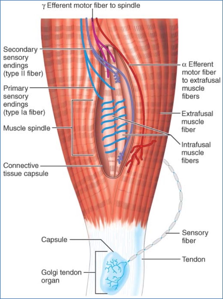
Muscle Spindles
- Location: Within skeletal muscle, among regular (extrafusal) fibres
- Structure: Encapsulated receptors containing intrafusal fibres
- Sensory Innervation:
- Central Region (non-contractile):
- Wrapped by type Ia fibres → detect rate and degree of stretch
- Spindle Ends:
- Wrapped by type II fibres → detect degree of stretch only
- Central Region (non-contractile):
- Motor Innervation:
- Gamma (γ) motor neurons → innervate distal contractile ends
- Maintain spindle sensitivity via γ-motor activity
α-γ Coactivation
- During voluntary contraction, α-motor neurons activate extrafusal fibres
- Simultaneously, γ-motor neurons activate intrafusal fibres
- Prevents spindles from becoming slack → maintains sensory output
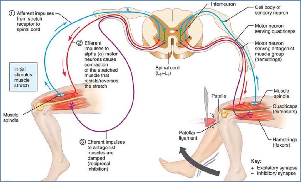
Stretch Reflex (Myotatic Reflex)
- Muscle stretch activates spindle
- Ia and II afferents send impulses to spinal cord
- Synapse directly with α-motor neurons
- Stimulation of the same muscle causes contraction
- Prevents overstretching
- Reciprocal inhibition: Inhibits antagonistic muscles
Example: Patellar (knee-jerk) reflex
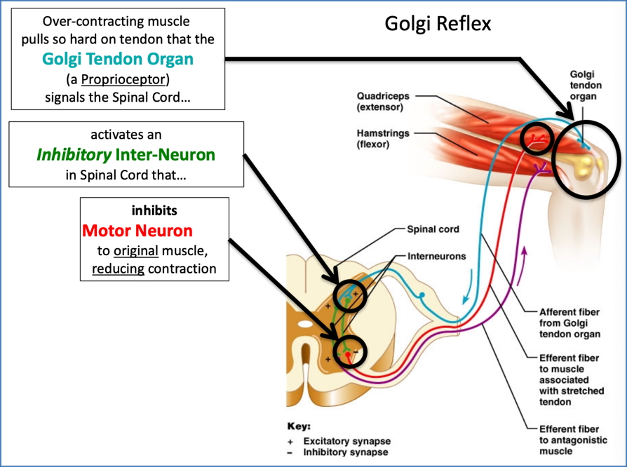
Golgi Tendon Organs (GTOs)
- Location: Within tendons near muscle–bone junction
- Innervation: Type Ib afferent fibres
- Function: Monitor muscle tension
Golgi Tendon Reflex
- Excessive tension → GTOs activated
- Type Ib afferents relay info to spinal cord
- Inhibits the contracting muscle (autogenic inhibition)
- Stimulates antagonist muscle
- Protective → prevents tendon rupture

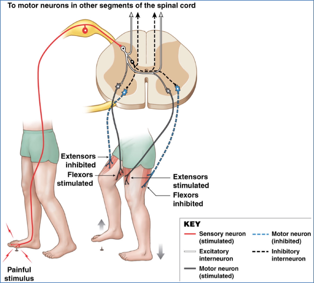
Flexor (Withdrawal) Reflex
- Triggered by painful cutaneous stimuli
- Polysynaptic → involves multiple interneurons
- Causes withdrawal of affected limb
- Coordinates contraction and relaxation of multiple muscles
- Example: Stepping on something sharp → leg withdrawal

Crossed Extensor Reflex
- Occurs in tandem with the flexor reflex
- Painful stimulus → affected limb withdraws
- Simultaneously, contralateral limb extends for support
- Mediated by interneurons crossing to opposite side of spinal cord
- Crucial for balance during withdrawal
Summary – Muscle Reflexes
Muscle reflexes provide rapid protective and postural responses to changes in muscle length, tension, and painful stimuli. Key players include muscle spindles (stretch reflex) and Golgi tendon organs (tension reflex), which contribute to proprioception and neuromuscular control. These reflex arcs are fundamental to both everyday movement and clinical neurological assessment. For a broader context, see our Musculoskeletal Overview page.