Table of Contents
Overview – The Axial Skeleton
The axial skeleton forms the central core of the body, comprising the skull, vertebral column, and thoracic cage. Its primary function is to protect vital structures such as the brain, spinal cord, and thoracic organs, while also supporting posture and movement. A critical component of this system is the vertebral column, which provides structural support and flexibility.
The Vertebral Column
General Features
- 33 vertebrae arranged into 5 regions:
- 7 Cervical
- 12 Thoracic
- 5 Lumbar
- 5 Sacral (fused)
- 4 Coccygeal (typically fused)
- Increases in vertebral size caudally due to greater load-bearing demands
- Protects the spinal cord
- Features fibrocartilaginous intervertebral discs for shock absorption
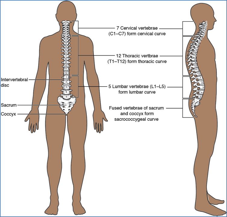
Curvatures of the Spine
- Primary Curvatures (concave anteriorly):
- Thoracic and sacral
- Secondary Curvatures (concave posteriorly):
- Cervical and lumbar
Abnormal Curvatures:
- Kyphosis – Exaggerated thoracic curve
- Lordosis – Exaggerated lumbar curve
- Scoliosis – Lateral curvature

Anatomy of a Typical Vertebra
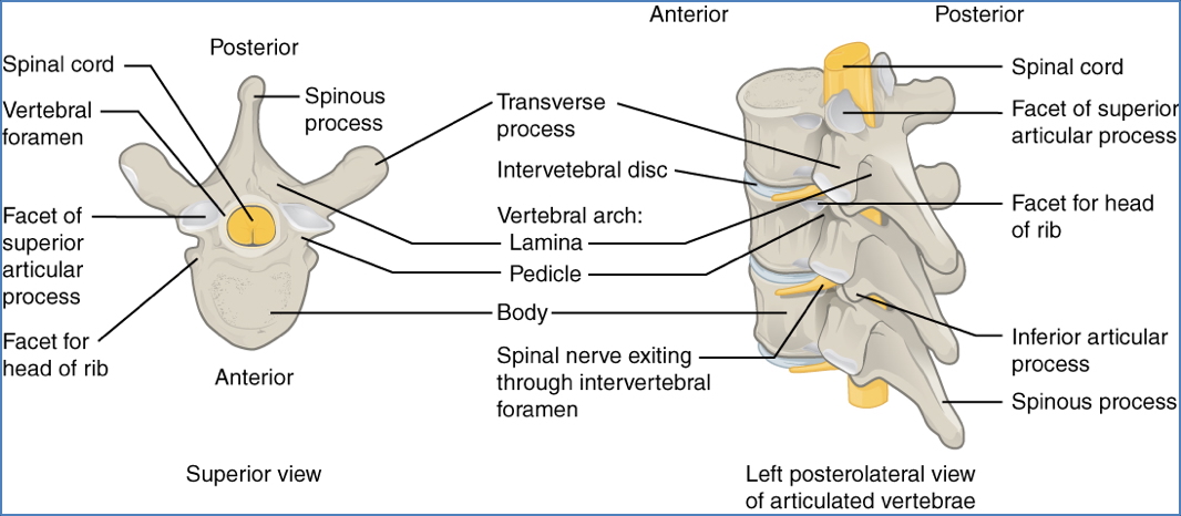
- Body – Weight-bearing anterior portion
- Vertebral Arch – Encloses the vertebral foramen
- Vertebral Foramen – Houses spinal cord
- Processes:
- Spinous Process
- Transverse Processes
- Superior & Inferior Articular Processes
- Intervertebral Foramina – Allow spinal nerves to exit the vertebral canal

Regional Vertebrae Characteristics
Cervical Vertebrae (C1–C7)
- Small body
- Large vertebral foramen
- Transverse foramina – Transmit vertebral arteries
- Bifid spinous processes (except C1 & C7)

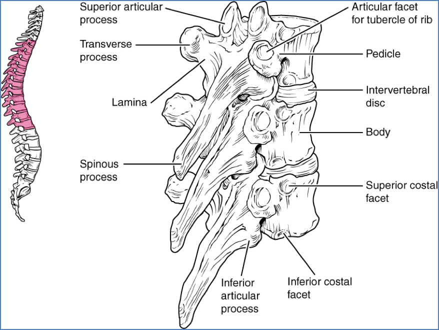
Thoracic Vertebrae (T1–T12)
- Medium-sized body
- Long spinous process
- Costal facets on transverse processes and vertebral bodies (articulate with ribs)
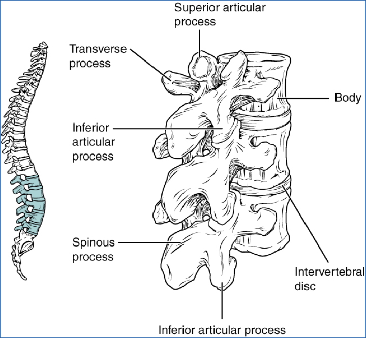
Lumbar Vertebrae (L1–L5)
- Very large body
- Short, blunt spinous processes
- No costal facets
- Smaller vertebral foramen
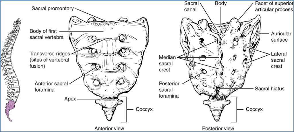
Sacrum (5 fused)
- Triangular shape
- Articulates with the ilium (sacroiliac joints)
Coccyx (3–5 fused)
- Remnant of a tail
- Angled anteriorly
Atypical Vertebrae
C1 – Atlas
- No body
- Ring of bone: anterior and posterior arches
- Transverse foramina
- Transverse ligament holds dens of C2 in place
- Supports the skull (atlanto-occipital joint)
C2 – Axis
- Prominent dens (odontoid process) projects superiorly
- Acts as a pivot for head rotation
- Also contains transverse foramina

Joints of the Vertebral Column
Atlanto-Occipital Joint
- Synovial ellipsoid (“egg & spoon”)
- Between skull and atlas
- Permits nodding (“yes”) motion
Atlanto-Axial Joint
- Combination of synovial pivot and planar joints
- Between atlas and axis
- Permits rotation (“no”) motion

Zygapophyseal (Facet) Joints
- Synovial planar joints
- Between articular processes of adjacent vertebrae
- Allow gliding movements and determine spinal flexibility

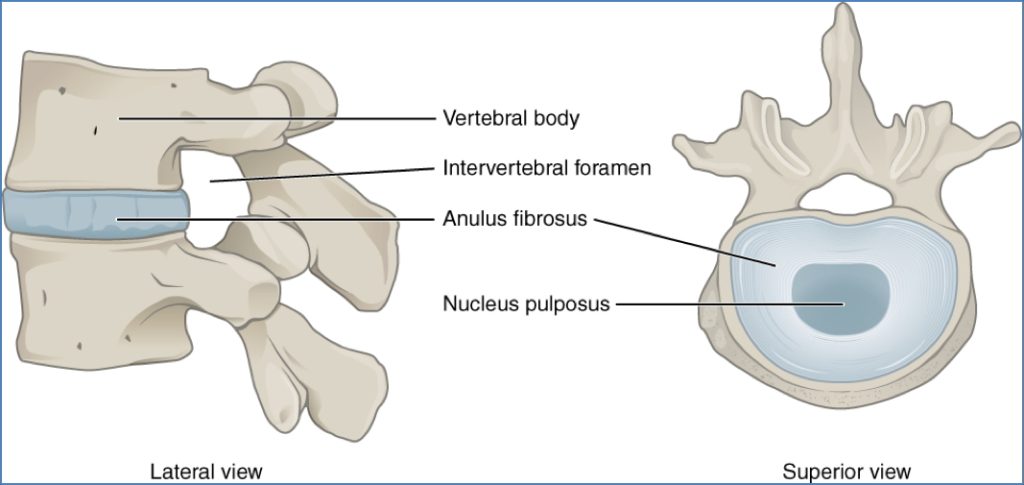
Intervertebral Discs
- Cartilaginous joints (symphyses)
- Permit limited movement and absorb shock
- Consist of:
- Annulus fibrosus: Outer fibrocartilage rings
- Nucleus pulposus: Inner gel-like core
- Disc herniation occurs when the nucleus protrudes through the annulus, possibly compressing nearby spinal nerves
Summary – The Axial Skeleton
The axial skeleton, anchored by the vertebral column, is essential for posture, support, and protection of central nervous system structures. Understanding vertebral regions, curvature types, and joint mechanics is fundamental to diagnosing spinal disorders and guiding treatments. For a broader context, see our Musculoskeletal Overview page.