Table of Contents
Overview – Cranial Nerves
This page provides a high-yield summary of the twelve cranial nerves — covering their names, functions, cell body locations, anatomical courses, and clinical associations. It’s designed to be a rapid, exam-focused reference for final-year medical students. For structural organisation, functional components, nuclei, and ganglia, see our Cranial Nerves Overview page.

I. Olfactory Nerve
- Type: Special Sensory (SVA – smell)
- Cell Body: Olfactory epithelium
- Course: Arises from olfactory mucosa → passes through cribriform plate of the ethmoid → synapses in olfactory bulb → projects via olfactory tract to primary olfactory cortex
- Function: Sense of smell
- Clinical: Anosmia (e.g. due to skull base fracture or viral infection)

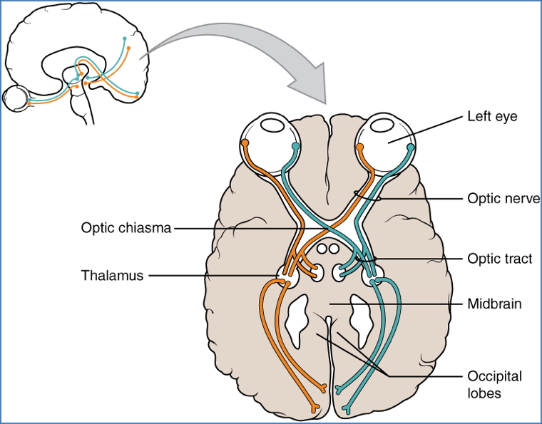
II. Optic Nerve
- Type: Special Sensory (SSA – vision)
- Cell Body: Retinal ganglion cells
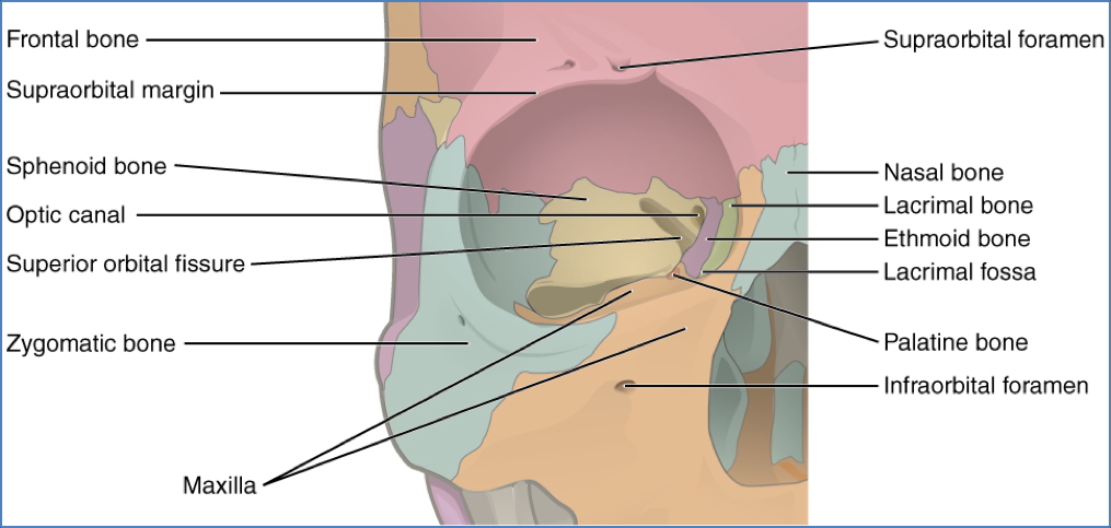
- Course: Arises from retina → exits via optic canal → forms optic chiasm (partial decussation) → continues as optic tract → synapses in thalamus → visual cortex
- Function: Vision and visual reflexes
- Clinical: Visual field loss (e.g. optic neuritis, stroke, tumours)
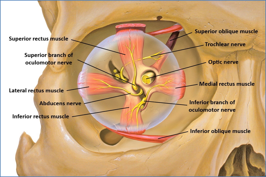
III. Oculomotor Nerve
- Type: Somatic Motor (GSE), Visceral Motor (GVE – parasympathetic)
- Cell Bodies:
- Somatic: Midbrain
- Parasympathetic: Presynaptic in midbrain, postsynaptic in ciliary ganglion
- Course: Emerges from midbrain → passes through superior orbital fissure → divides into superior/inferior branches to extraocular muscles
- Function:
- Eye movement (superior, medial, inferior recti + inferior oblique)
- Levator palpebrae (eyelid elevation)
- Parasympathetic to pupil sphincter and ciliary muscle
- Clinical: Ptosis, “down and out” gaze, dilated pupil
IV. Trochlear Nerve
- Type: Somatic Motor (GSE)
- Cell Body: Midbrain
- Course: Emerges dorsally from midbrain → passes through superior orbital fissure → innervates superior oblique
- Function: Depresses and intorts eye (down & in)
- Clinical: Vertical diplopia, worsened with looking down (e.g. stairs)
V. Trigeminal Nerve
V1 – Ophthalmic
- Type: General Sensory (GSA)
- Cell Body: Trigeminal ganglion
- Course: Enters skull via superior orbital fissure
- Function: Sensation from forehead, scalp, cornea, upper eyelid
- Clinical: Loss of corneal reflex (afferent limb)
V2 – Maxillary
- Type: General Sensory (GSA)
- Cell Body: Trigeminal ganglion
- Course: Enters skull via foramen rotundum
- Function: Sensation from midface, upper teeth, sinuses, palate
V3 – Mandibular
- Type: General Sensory + Branchial Motor (SVE)
- Cell Bodies: Trigeminal ganglion (sensory), motor nucleus of V (motor)
- Course: Enters via foramen ovale
- Function:
- Sensation from lower face, teeth, tongue
- Motor to muscles of mastication, mylohyoid, anterior digastric
- Clinical: Trigeminal neuralgia, jaw deviation

VI. Abducens Nerve
- Type: Somatic Motor (GSE)
- Cell Body: Pons
- Course: Arises from pons → passes through superior orbital fissure → innervates lateral rectus
- Function: Abducts the eye
- Clinical: Diplopia on lateral gaze (medial deviation)
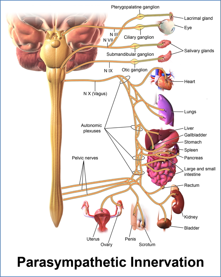
VII. Facial Nerve
- Type: Branchial Motor (SVE), Visceral Motor (GVE), Special Sensory (SVA), General Sensory (GSA)
- Cell Bodies:
- Motor: Pons
- Parasymp: Pons → pterygopalatine / submandibular ganglia
- Sensory: Geniculate ganglion
- Course: Exits pons → enters internal acoustic meatus → traverses facial canal → exits skull via stylomastoid foramen
- Function:
- Facial expression muscles
- Taste (ant. 2/3 tongue)
- Parasympathetic to lacrimal, submandibular, sublingual glands
- Clinical: Bell’s palsy, loss of taste, dry eyes/mouth

VIII. Vestibulocochlear Nerve
- Type: Special Sensory (SSA)
- Cell Bodies:
- Vestibular: Vestibular ganglion
- Cochlear: Spiral ganglion
- Course: Arises from inner ear → enters brainstem at pontomedullary junction via internal acoustic meatus
- Function:
- Hearing (cochlear)
- Balance (vestibular)
- Clinical: Sensorineural hearing loss, vertigo, nystagmus

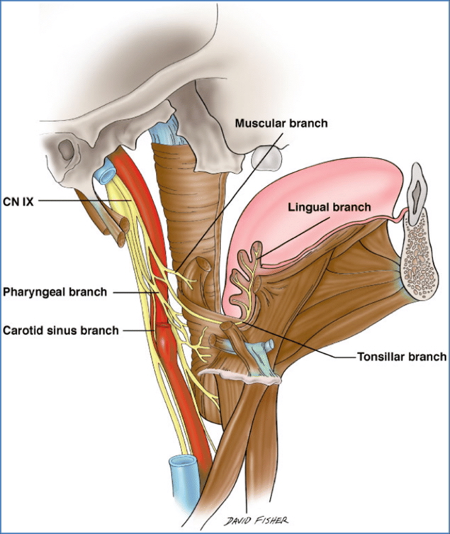
IX. Glossopharyngeal Nerve
- Type: SVE, GVE, GVA, SVA, GSA
- Cell Bodies:
- Motor: Medulla
- Parasymp: Otic ganglion
- Sensory: Superior & inferior ganglia
- Course: Emerges from medulla → exits via jugular foramen
- Function:
- Stylopharyngeus (swallowing)
- Parotid gland secretion
- Taste + sensation (post. 1/3 tongue)
- Carotid body/sinus inputs
- Clinical: Loss of gag reflex, BP dysregulation
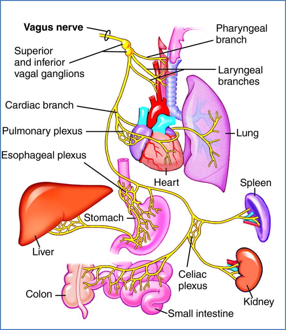
X. Vagus Nerve
- Type: SVE, GVE, GVA, SVA, GSA
- Cell Bodies:
- Motor: Medulla
- Parasymp: Postsynaptic in thoracic/abdominal ganglia
- Sensory: Superior & inferior ganglia
- Course: Emerges from medulla → exits via jugular foramen → travels through neck, thorax, abdomen
- Function:
- Swallowing, speech (pharynx, larynx)
- Parasympathetic to thoracoabdominal viscera
- Taste (epiglottis)
- Clinical: Hoarseness, uvula deviation, dysphagia
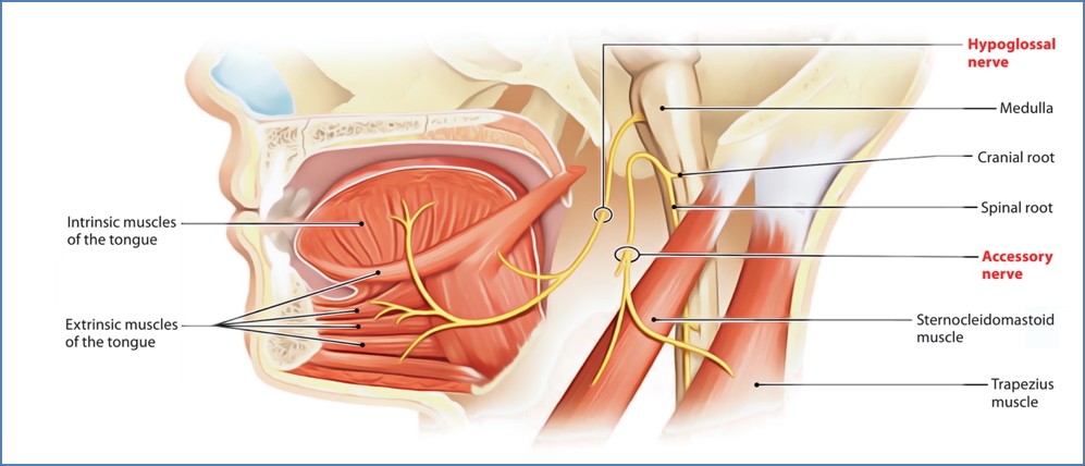
XI. Accessory Nerve
- Type: Somatic Motor (GSE)
- Cell Body: Spinal cord (C1–C5)
- Course: Enters skull via foramen magnum, exits via jugular foramen
- Function: SCM + trapezius control
- Clinical: Shoulder droop, weak head rotation
XII. Hypoglossal Nerve
- Type: Somatic Motor (GSE)
- Cell Body: Medulla
- Course: Arises from medulla → exits via hypoglossal canal → travels to intrinsic + extrinsic tongue muscles
- Function: Tongue movement (speech, swallowing, bolus control)
- Clinical: Tongue deviation (toward lesion side)
Summary – Cranial Nerves
This page consolidates the names, functions, pathways, and clinical associations of all twelve cranial nerves — with detailed anatomical and embryological context. These nerves are essential for sensation, movement, reflexes, and autonomic control in the head, neck, and thorax. For developmental origins and ganglia, see our Cranial Nerves Overview page.