Table of Contents
Overview – Vision
Vision is the dominant human sense and central to clinical examination, navigation, and communication. Understanding the anatomy and physiology of the eye is crucial in both clinical diagnosis (e.g. red eye, pupil abnormalities, visual field loss) and neurological localisation. This page covers the structural components of the eye — from protective structures to photoreceptors — and walks through the visual processing pathway from photon capture to cortical perception.
Accessory Structures of the Eye
- Eyebrows:
- Shade eyes
- Divert sweat away from eyes
- Eyelids (Palpebrae):
- Protect from foreign bodies
- Blinking prevents dryness
- Conjunctiva:
- Transparent mucous membrane (palpebral + bulbar)
- Secretes lubricating mucus

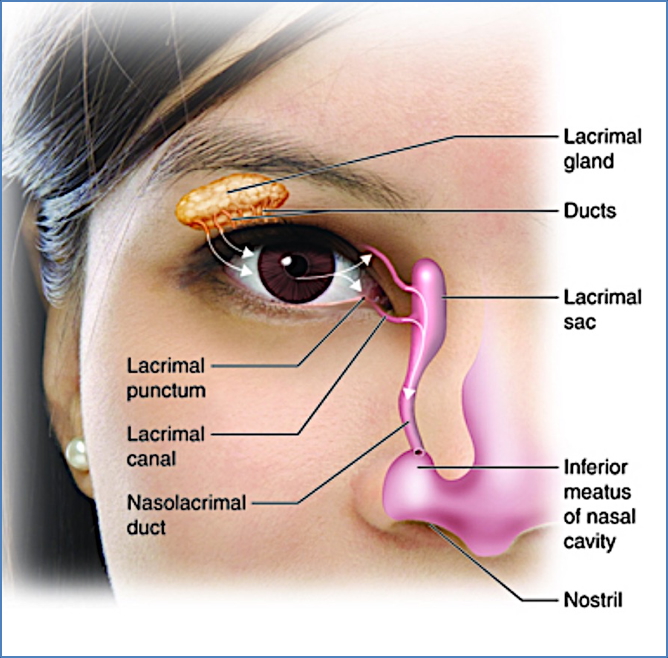
- Lacrimal Apparatus:
- Lacrimal gland (tear production)
- Lacrimal puncta → canaliculi → lacrimal sac → nasolacrimal duct
- Tears contain mucus, antibodies, lysozyme
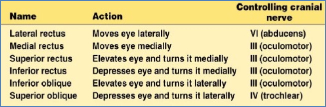
- Extrinsic Eye Muscles:
- 6 muscles move the eye
- 4 rectus from annular ring
- 2 oblique muscles refine vertical movement


Eye Anatomy
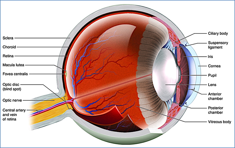
Layers of the Eye Wall
- Fibrous Layer (Outer)
- Sclera: Tough, white connective tissue; continuous with dura
- Cornea: Transparent; main refractive surface of the eye
- Vascular Layer (Middle)
- Choroid: Pigmented, vascular; nourishes outer retina
- Ciliary Body:
- Smooth muscle ring
- Adjusts lens shape via ciliary zonule
- Iris:
- Coloured diaphragm with central pupil
- Muscles:
- Sphincter pupillae → constriction
- Dilator pupillae → dilation
- Retinal Layer (Inner)
- Pigmented Layer:
- Light absorption
- Phagocytosis & vitamin A storage
- Neural Layer:
- Photoreceptors (rods & cones)
- Bipolar neurons
- Ganglion cells (form optic nerve)
- Amacrine + horizontal cells = local processing
- Key Landmarks:
- Optic Disc (blind spot)
- Macula lutea: Cone-rich
- Fovea centralis: Only cones; sharpest vision
- Pigmented Layer:

Segments & Fluids of the Eye
Anterior Segment
- Anterior to lens
- Aqueous humour:
- Formed by ciliary processes
- Flows posterior → anterior chamber
- Drains via Canal of Schlemm
- Functions:
- Nourishes cornea/lens
- Maintains intraocular pressure

Posterior Segment
- Posterior to lens
- Vitreous humour:
- Formed during embryogenesis
- Functions:
- Light transmission
- Supports retina
- Maintains intraocular pressure
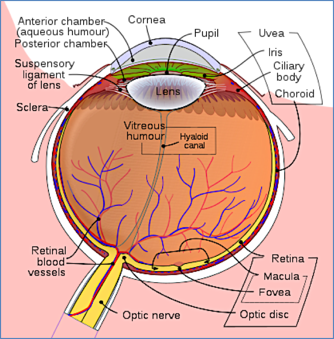
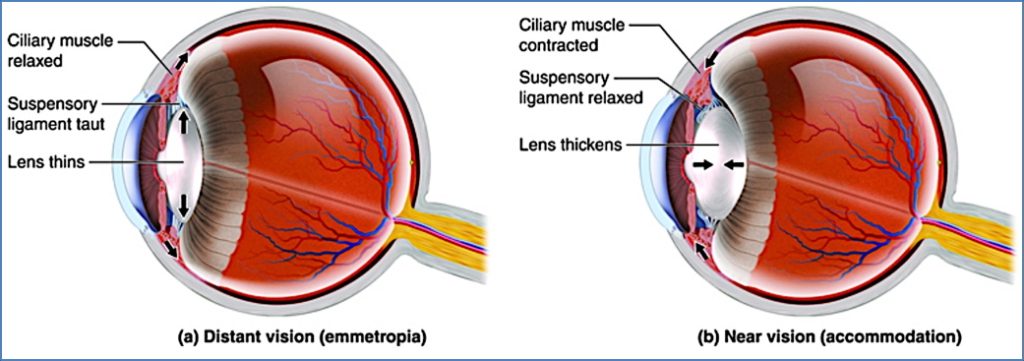
The Lens
- Biconvex, transparent, flexible
- Held by ciliary zonule
- Composed of:
- Lens epithelium (anterior)
- Lens fibres (bulk, layered)
- Accommodation:
- Adjusts focal power to focus light onto retina
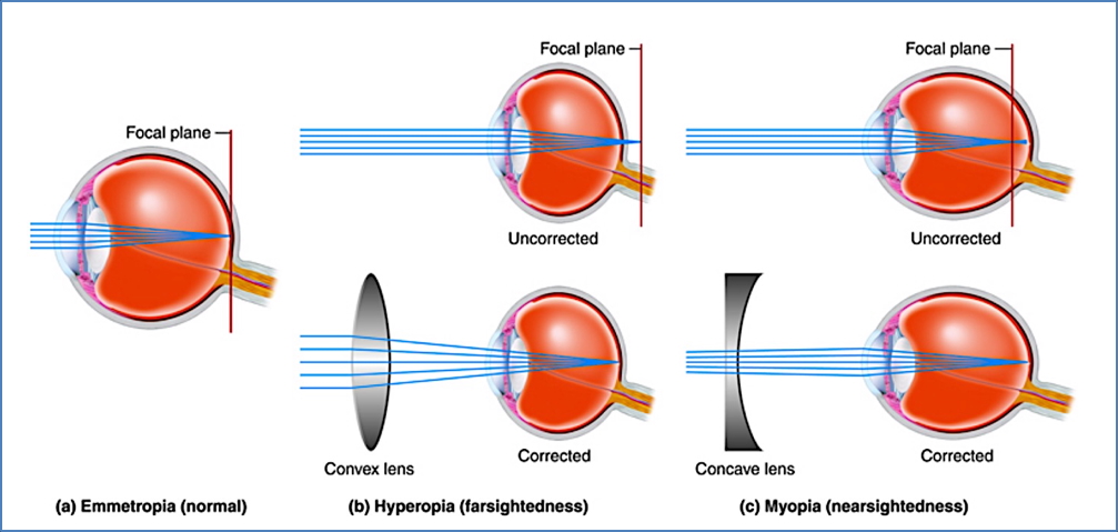
- Focal Disorders:
- Emmetropia = Normal
- Myopia = Eye too long
- Hyperopia = Eye too short
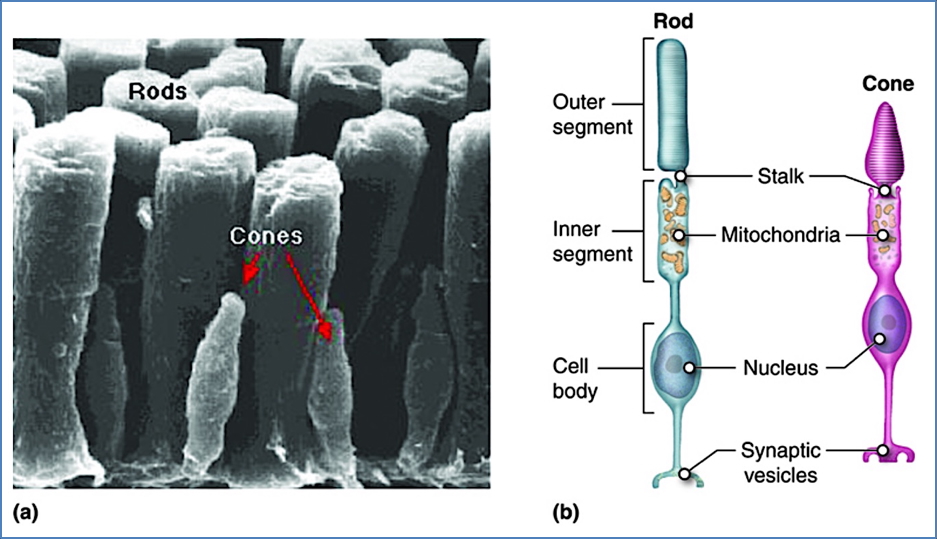
Photoreceptors
- Convert light into neural signals
- Rod Cells
- High sensitivity
- Monochrome, dim-light vision
- Low spatial resolution
- Cone Cells
- Colour vision
- High resolution
- Require bright light
- 1:1 ganglion connections → sharp image
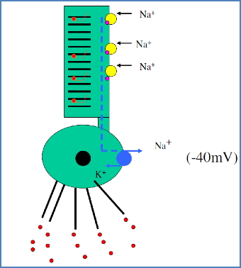
Phototransduction
- Retinal (vitamin A derivative) + Opsins = Photopigments
- Dark (Inactive) State:
- cGMP keeps Na⁺ channels open → depolarised (-40 mV)
- Glutamate released → bipolar cells inhibited
- Light Activation:
- Retinal isomerises (11-cis → all-trans)
- cGMP breaks down → Na⁺ channels close
- Repolarisation (-70 mV) → glutamate release stops
- Bipolar cells activated → ganglion APs → optic nerve
Light and Dark Adaptation
- Light Adaptation:
- Rod system shuts off
- Cones take over
- Takes up to 60 seconds
- Dark Adaptation:
- Cones off, rods bleached
- Rhodopsin regenerates → rod function returns
- May take 30+ minutes
Retinal Signal Processing
- Direct Path:
- Photoreceptor → Bipolar Cell → Ganglion Cell
- Horizontal/Amacrine Cells → Lateral inhibition & contrast enhancement
Visual Pathway
- Ganglion axons = Optic nerve
- Optic chiasm: Partial decussation
- Optic tract →
- Superior colliculi (midbrain): Reflex eye movements
- Lateral geniculate nucleus (thalamus) →
- Optic radiation → Primary visual cortex (occipital lobe)
- Higher order processing in temporal, parietal, and frontal lobes

Summary – Vision
Vision integrates the structural complexity of the eye with precise neural mechanisms of transduction, processing, and cortical interpretation. From accessory structures to the retina and beyond, every component plays a critical role in directing photons into meaningful perception.
For a broader context, see our Nervous System Overview page.