Table of Contents
Overview – Renal Stones
Renal stones (nephrolithiasis or urolithiasis) are crystalline mineral deposits that form in the urinary tract due to supersaturation of solutes in the urine. They are a common cause of flank pain, haematuria, and potential urinary obstruction. While many stones pass spontaneously, some require medical or surgical intervention. Early identification and prevention are key to reducing recurrence and complications such as post-renal failure.
Definition
- Nephrolithiasis refers to stones formed in the kidney.
- Urolithiasis refers to stones located anywhere along the urinary tract (e.g. ureters, bladder).
Aetiology
- Hypercalcaemia (≈80%)
- Causes: hyperparathyroidism, high calcium intake
- → Calcium oxalate/phosphate stones
- Chronic UTI (≈15%)
- Urease-producing organisms (e.g. Proteus, Pseudomonas, Klebsiella)
- → Triple phosphate/struvite/staghorn calculi
- Uraemia / Gout
- → Urate stones
- Other rare causes:
- Cystine stones
- Xanthine stones
Pathophysiology
- Supersaturation of urine → solute precipitation → crystal aggregation → stone formation
- Specific mechanisms:
- Hypercalcaemia → excess urinary calcium precipitates
- Chronic UTI → ammonia production increases urine pH → struvite crystallisation
- Uric acid binds sodium → forms monosodium urate crystals
- Stones can obstruct urine flow → hydronephrosis → stretching of renal capsule → pain
Risk Factors
- Family history
- Recurrent UTIs
- Neurogenic bladder or vesicoureteral reflux
- Congenital abnormalities (e.g. horseshoe kidney)
- Hyperuricaemia:
- High-purine diet (red meat, shellfish)
- High cell turnover (e.g. leukaemia, chemotherapy)
- Hypercalcaemia (e.g. inflammatory bowel disease, calcium supplements)
- Obesity, high salt intake
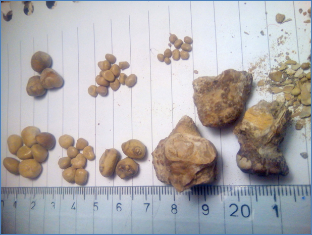
Morphology
Calcium Stones (~80%)
- Small (1–3 mm), hard, sharp-edged
- Radio-opaque on X-ray
Struvite/Staghorn Stones (~15%)
- Large, moulded to renal pelvis/calyces (“staghorn”)
- Associated with squamous metaplasia
- Often due to chronic infection
Urate Stones
- Pebble-like, rough outer surface
- Softer core
- Radiolucent on X-ray
Clinical Features
- Usually unilateral
- Painful haematuria (macro- or microscopic)
- Severe, colicky flank pain – patient often writhes in distress
- Stone-specific locations and symptoms:
- Ureteropelvic junction: deep flank pain without radiation
- Mid-ureter: colicky loin-to-groin pain, possibly radiating to testes/labia
- Ureterovesical junction: dysuria, urinary frequency, pain at tip of penis
Complications
- Hydronephrosis
- Post-renal failure
- Infection (UTI, pyelonephritis, perinephric abscess)
Investigations
- Abdominal ultrasound:
- Preferred in pregnancy; detects hydronephrosis and stones
- Abdominal X-ray (AXR):
- Detects radio-opaque stones (e.g. calcium)
- Non-contrast CT-KUB:
- Best for identifying size, location, and composition
- UECs:
- Hypercalcaemia or uraemia
- Urinalysis:
- Haematuria
- Crystals in urine
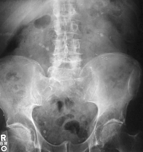
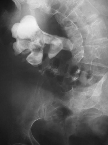
Nevit Dilmen, CC BY-SA 3.0 <https://creativecommons.org/licenses/by-sa/3.0>, via Wikimedia Commons
Management
- Supportive care:
- Analgesia (e.g. NSAIDs)
- Hydration
- Most stones <5 mm pass spontaneously
- Medical management:
- Urine alkalinisers (Na-bicarbonate, K-citrate) → dissolve urate stones
- Alpha blockers or calcium channel blockers → reduce ureteric spasm
- Extracorporeal shock wave lithotripsy (ESWL):
- Non-invasive stone fragmentation (best for calcium stones)
- Surgical options:
- Ureteroscopy, stenting, or percutaneous nephrolithotomy
- Indicated for large or non-passing stones
Summary – Renal Stones
Renal stones are a common urological condition caused by solute precipitation in the urinary tract. Calcium stones are most frequent, followed by struvite and urate stones. Clinical features include flank pain and haematuria, and management ranges from supportive care to surgical removal. For a broader context, see our Renal Overview page.