Table of Contents
Overview – Bronchiectasis
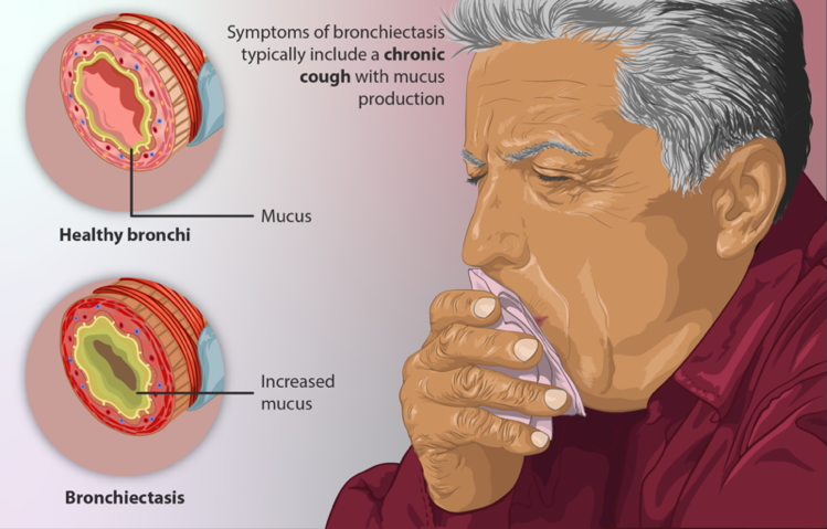
Bronchiectasis is a chronic respiratory condition characterized by irreversible dilation of the bronchi due to repeated inflammation and infection. It results from structural damage to the airway walls, typically following necrotizing infections or conditions that impair mucociliary clearance. Patients often present with chronic cough and copious, purulent sputum. Understanding its pathogenesis, common causes (like cystic fibrosis), and complications is essential in both respiratory and infectious disease practice.
Definition
Bronchiectasis is a localized, permanent dilation of part of the bronchial tree due to destruction of the muscle and elastic tissue supporting the airways.
Aetiology
Bronchiectasis often results from recurrent or severe infections that damage the bronchial walls. Common underlying causes include:
- Cystic Fibrosis – most common cause in children
- Post-infectious complications – e.g., measles, pertussis, tuberculosis
- Bronchial obstruction – tumors or foreign bodies
- Immunodeficiencies
- HIV infection
- Primary ciliary dyskinesia (e.g. Kartagener’s syndrome)
Pathogenesis
- Recurrent or severe bacterial infections lead to:
- Chronic bronchial inflammation
- Destruction of smooth muscle and elastic tissue in airway walls
- Resulting fibrosis and structural weakening cause:
- Irregular, permanent airway dilation
- Accumulation of pus and mucus
- Progressive airway damage and impaired clearance

Morphology
- Most commonly affects the lower lobes
- Bronchi may be dilated up to 4× normal size, often extending to the pleura
- Radiological findings:
- CXR: Bronchial markings visible >1/3 across the lung field
- CT chest (gold standard): “Tram-track” or “signet ring” appearance
Clinical Features
Symptoms
- Chronic productive cough
- Copious purulent sputum (green or yellow)
- Haemoptysis (variable)
- Recurrent respiratory infections
- Fatigue, breathlessness, low-grade fever
Complications
- Recurrent pneumonias (Staph, Strep, Pseudomonas, etc.)
- Empyema
- Septicaemia
- Meningitis (rare, due to hematogenous spread)
- Respiratory failure (in advanced cases)
Investigations
- Chest X-Ray: Prominent peribronchial markings; signs of volume loss
- High-Resolution CT (HRCT): Diagnostic modality of choice
- FBC: To assess for signs of acute infection
- Sputum culture: Identify bacterial pathogens (esp. Pseudomonas)
- Spirometry: May show obstructive or mixed pattern
Management
Supportive
- Physiotherapy – postural drainage, chest percussion (cupping), breathing exercises
- Hydration – to help loosen mucus
Medical
- Antibiotics – indicated during exacerbations or based on sputum cultures
- Tobramycin (for Pseudomonas)
- Amoxicillin-clavulanate, macrolides (empirical)
Preventive
- Vaccinations – influenza, pneumococcal
- Smoking cessation
Surgical (rare)
- Considered in localized disease unresponsive to medical therapy
Summary – Bronchiectasis
Bronchiectasis is a chronic lung condition marked by irreversible bronchial dilation due to structural damage from recurrent infections. Hallmark symptoms include persistent productive cough and purulent sputum. Management focuses on airway clearance, infection control, and addressing underlying causes such as cystic fibrosis. For broader context, visit our Respiratory Overview page.