Table of Contents
Overview – Upper Airway Anatomy
The upper airways conduct, warm, humidify, and filter inspired air. This region includes the nose, nasal cavity, sinuses, pharynx, and associated skeletal structures. Understanding upper airway anatomy is essential for clinical ENT, respiration, and head & neck anatomy exams.
Structural Divisions
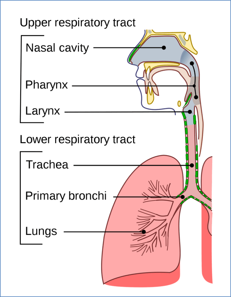
- Upper airways (conducting zone):
- Nose → trachea
- Functions: filters particulate matter, warms and moistens air through mucosal epithelium
- Lower airways (respiratory zone):
- Bronchi → lungs
- Functions: gas exchange (oxygen uptake, carbon dioxide removal)
Facial Skeleton and Connections
- Communication routes:
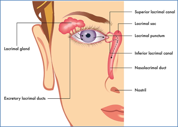
- Eye orbits ↔ nasal cavities (nasolacrimal duct)
- Nasal cavities ↔ paranasal sinuses
- Nasal cavities ↔ oral cavity
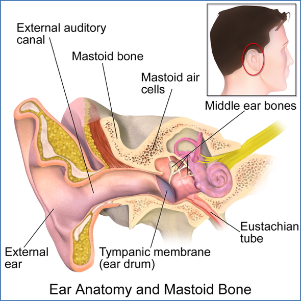
- Ears ↔ pharynx (Eustachian tube for pressure equalisation)
- Pharynx ↔ larynx
- Maxillae (paired): fused medially, carry upper teeth, form anterior 2/3 of hard palate, articulate with all facial bones except mandible
- Frontal bone: anterior cranium, contains frontal sinuses, articulates with ethmoid
- Nasal bones: form nasal bridge, support external cartilage

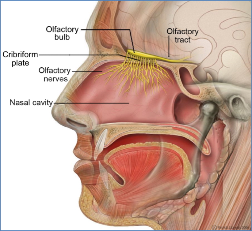
- Ethmoid bone: forms much of nasal cavity, anchors nasal cartilage, turbinates air
- Cribriform plates (olfactory foramina)
- Crista galli (anchors brain dura)
- Perpendicular plate (superior nasal septum)
- Lateral masses (ethmoid sinuses)
- Superior and middle nasal conchae (air filtration, warming, humidification)
- Inferior nasal conchae: separate paired scroll-like bones, attached to maxilla, add turbulence
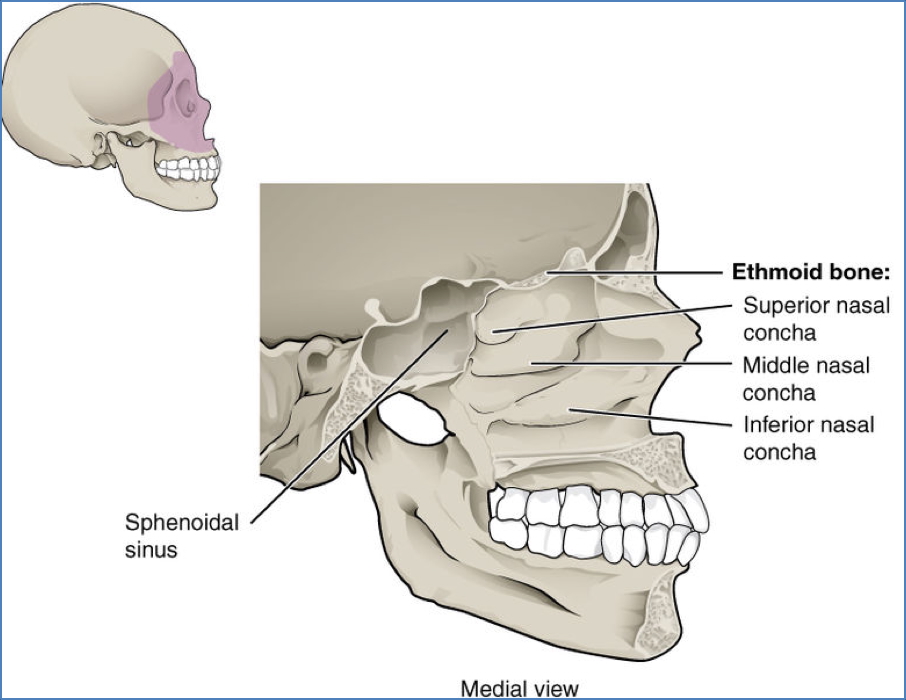

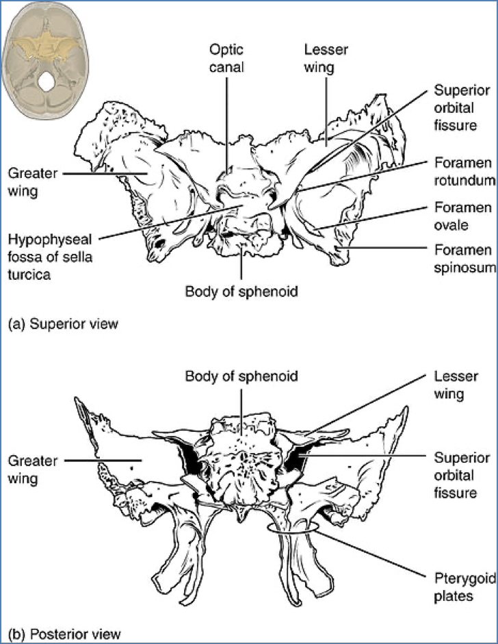
- Sphenoid bone: keystone of cranium, butterfly-shaped, contains paired sphenoid sinuses
The Nose
- Functions: respiration, warms and moistens air, filters debris, resonance chamber, houses olfactory receptors
- External nose: nasal and frontal bones (superior), maxilla (lateral), cartilage (inferior)
- Nasal cavity:
- Entry via nostrils lined with hair (vibrissae), sebaceous and sweat glands
- Olfactory mucosa: specialised epithelium, olfactory neurons synapse with cranial nerve I
- Respiratory mucosa: pseudostratified columnar epithelium with cilia and goblet cells
- Nasal septum: formed by perpendicular plate of ethmoid (upper 2/3) and vomer (lower 1/3)
- Roof: ethmoid and sphenoid bones
- Floor: hard palate (palatine bone), soft palate (uvula)
- Lateral walls: superior, middle, inferior conchae → ↑ surface area, turbulence, moisture/heat exchange
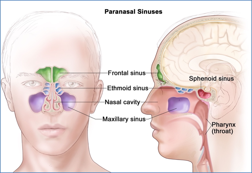
Paranasal Sinuses
- Located in: maxilla, frontal, sphenoid, ethmoid bones
- Continuous with nasal cavity
- Functions: increase mucosal surface area, humidify and warm air, lighten skull, enhance voice resonance
- Clinical note: blockage during infection → mucus buildup → sinus pain
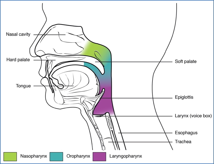
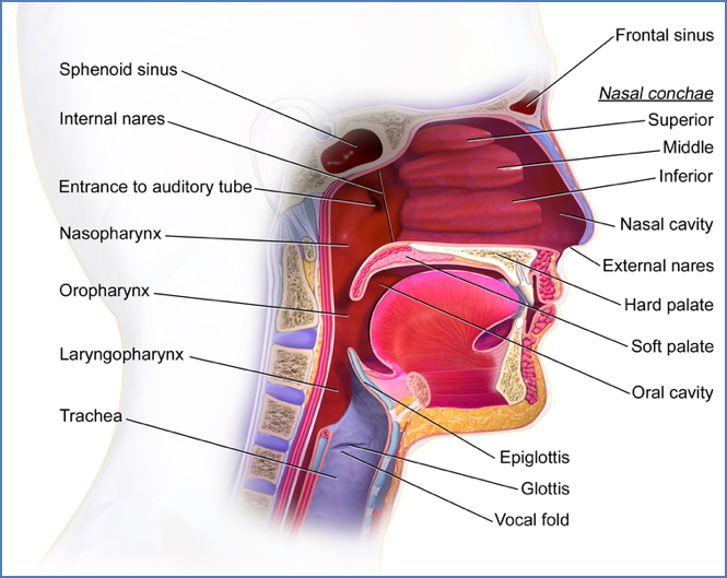
The Pharynx
- Connects nasal cavities, oral cavity, and oesophagus
- Regions and epithelium:
- Nasopharynx – transmits air only; pseudostratified ciliated epithelium
- Oropharynx – transmits food and air; stratified squamous epithelium
- Laryngopharynx – transmits food and air; stratified squamous epithelium; food passage takes priority during swallowing
- Muscles of pharynx:
- Constrictors (superior, middle, inferior): propel food towards laryngopharynx
- Longitudinal muscles (palatopharyngeus, salpingopharyngeus, stylopharyngeus): elevate pharynx and protect airway
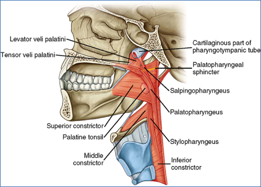
- Soft palate: separates oral cavity and nasopharynx, functions in swallowing
- Muscles: levator veli palatini, tensor veli palatini, palatoglossus, palatopharyngeus, musculus uvulae
- Innervation: vagus nerve
The Larynx
The larynx (“voicebox”) connects the pharynx to the trachea and is essential for airway protection and phonation.
- Position:
- Superior attachment to hyoid bone
- Inferior continuation with the trachea
- Functions:
- Maintains an open airway for breathing
- Directs food and air into appropriate channels
- Produces sound (phonation)
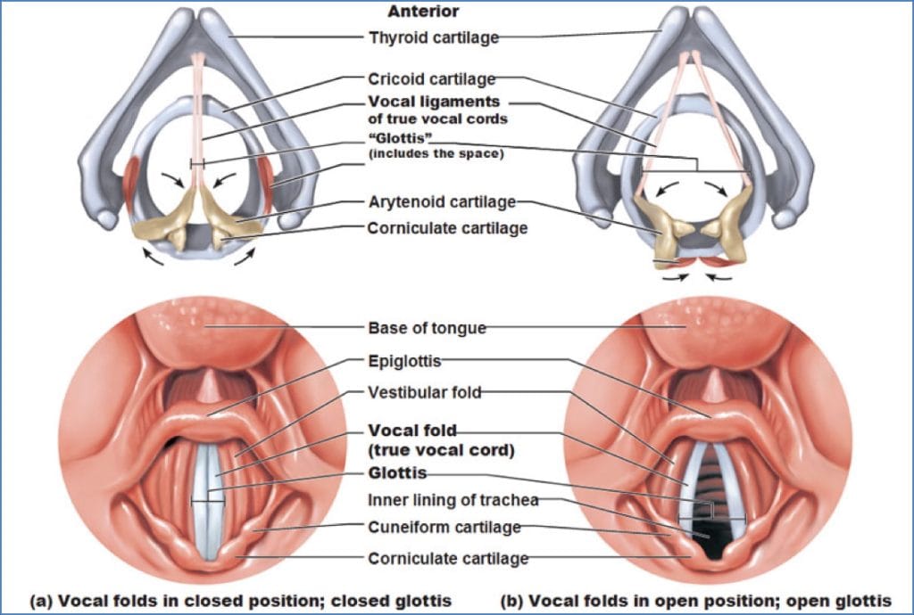
- Cartilages:
- Unpaired (3): Thyroid, Cricoid, Epiglottis → form tubular skeletal framework
- Paired (3 × 2): Arytenoid, Cuneiform, Corniculate → control vocal ligament movement

- Vocal ligaments (“true vocal cords”):
- Elastic fibres covered in mucosa
- Stretch between thyroid and arytenoid cartilages
- Vibrate as air passes → phonation (tension = pitch)
- White in colour due to lack of blood supply
- Vestibular folds (“false vocal cords”):
- Quadrangular membrane
- Do not produce sound
- Aid in closing glottis during swallowing
- Intrinsic laryngeal muscles (vagus nerve innervation):
- Functions: alter vocal cord length, tension, and position
- 2x Cricothyroid
- 2x Vocalis
- 2x Transverse arytenoids
- 2x Oblique arytenoids
- 2x Posterior crico-arytenoids
- 2x Lateral crico-arytenoids
- 2x Thyromuscularis

Review of Entire Respiratory Anatomy:

Summary – Upper Airway Anatomy
Upper airway anatomy includes the conducting portions of the respiratory tract, from the nose to the trachea, where air is filtered, humidified, and warmed. Structures such as the nasal cavity, paranasal sinuses, and pharynx play roles in respiration, olfaction, speech, and protection. Understanding these connections is crucial in clinical practice, particularly in airway management and sinus disease. For a broader context, see our Respiratory Overview page.